


 Español (pdf)
 Articulo en XML
 Referencias del artículo
 Enviar articulo por email
 Citado por SciELO
  Similares en
SciELO
Similares en
SciELO  uBio
uBio 
 Permalink
INTRODUCTION
According to the World Health Organization (WHO, 2016), congenital anomalies are also known as birth defects, congenital disorders or congenital malformations. Congenital defects can be defined as structural or functional abnormalities that occur during intrauterine life and can be identified prenatally, at birth or, sometimes, can only be detected later in the early postbirth stages. In simple terms, congenital refers to the existence on or before birth.
Initial embryonic development of the ducts, tubules, and external genitalia is the same in both sexes. The internal organs in this stage consist of two bilaterally symmetrical ductal systems - the paramesonephric (Müllerian; female) and mesonephric (Wolffian; male) ducts - and the gonads. Although the uterine tubes, uterine horns and body, cervix, and cranial vagina develop from the paramesonephric duct, the mesonephric tubes become the rete and contribute to the sex cords of the ovary. Generally, the ducts of the opposite sex retrogress (Schlafer and Foster, 2016).
The primitive cloaca (embryo and initial fetal stages) is first separated into the rectum and the urogenital sinus by the urorectal bend. Afterwards, the urogenital sinus differentiates into the urinary bladder and urethra. Failure of the urorectal fold to completely separate the primitive cloaca results in atresia ani (Vianna and Tobias, 2005).
CASE REPORT
In November 2018, the animal care workers at a ranch in Puerto Salgar, Colombia reported that a two-day-old Gyr calf had not been able to defecate. At the veterinary consultation, the inspection showed that the calf was in the agonizing state, had urinary and fecal tenesmus and at the clinical examination the anus and external reproductive organs were no presented, referring to a congenital atresia of the anus and agenesis of the vulva (Figure 1). Due to animal welfare conditions, it was decided to perform euthanasia and necropsy.
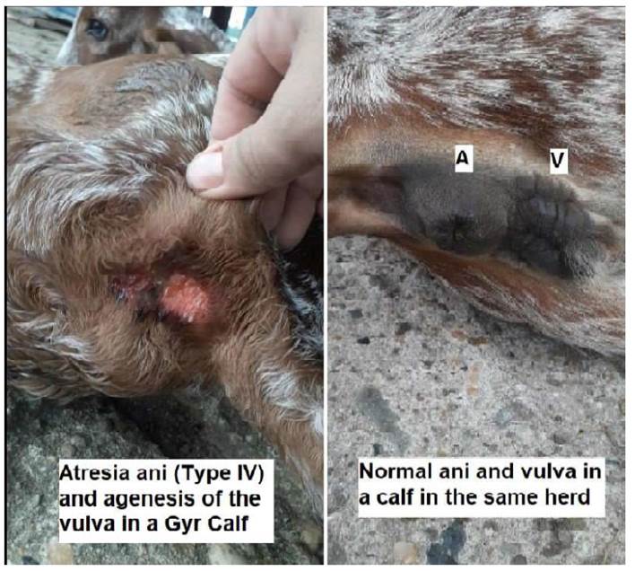
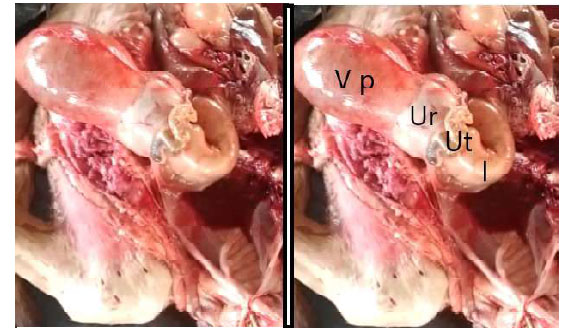
Figure 2 Plethoric bladder and urethra and rectovaginal fistula. V p: Plethoric bladder; Ur: Urethra; Ut: Uterine horns; I: intestines
In the necropsy was found congenital atresia ani (type IV), agenesis of the vulva, plethoric bladder and urethra (Figure 2), and dilated ureters due to vesicoureteral reflux, rectovaginal fistula, urosalpinx-urometrocolpos and coprosalpinx-coprometrocolpos.
DISCUSSION
Congenital defects refer to anomalies presented at birth derived from faults originated during gestation (Noden and deLahunta, 1985). The origin of these anomalies could be due to genetic causes or habitat influence and produce structure or function alterations which may be lethal, semilethal, or compatible with life (Ghanem et al., 2004). In many cases, the causes are unknown (Lombardero and Yllera, 2014). In this specific case, the cause is unknown and the defect was lethal, because if the calf could not excrete urine and feces naturally due to a lack of continuity of the digestive and urinary ducts, these systems would end collapsed as a result of a possible secondary hydronephrosis, rupture of bladder, ureters, and intestines, peritonitis, sepsis and death.
Agenesis of the vulva has been reported in canines (Meij et al., 1990; Jae-il et al., 2007), in lambs (Chaudhary et al., 2016), cattle (Wamaitha et al., 2015) and buffalo (Krishna et al., 2009). Failure of the differentiation of cloacal folds into anal and urogenital folds outcomes in the malformation of the anus and vagina (Noden and deLahunta, 1985). There have been reports of different digestive and reproductive congenital defect, including an unusual colon atresia in a calf (Lombardero and Yllera, 2014), atresia ani in a calf (Suthar et al., 2010). Atresia ani with rectovaginal fistula has been reported in 11 cases in various species, and atresia ani et vulvi was diagnosed in three native calves in India (Gamal, 2006). Besides, agenesis of the vulva with atresia ani-et-distal recti in a calf (Wamaitha et al., 2015) and atresia ani et recti along with pervious urachus in Sahiwal cow calves (Singh et al., 2019). This case described an atresia ani (Type IV), agenesis of the vulva and rectovaginal fistula in a Gyr calf.
Atresia ani has four anatomic variants (Bright and Bauer, 1994): «Type I: a membrane over the anal opening remains, with the rectum ending as a blind pouch just cranial to the closed anus; Type II: the anus is closed as in type I, but the rectal pouch is located somewhat cranial to the membrane overlying the anus; Type III: the rectum ends as a blind pouch cranially within the pelvic canal (rectal atresia), whereas the terminal rectum and anus are normal; Type IV: occurs in females and atresia ani exists with a persistent communication between the rectum and the vagina (rectovaginal fistula). This fistula can occur with a normal anal opening as well». Based on these definitions, the actual case reported is an atresia ani type IV.
Urosalpinx-metrocolpos and coprosalpinx-metrocolpos and dilated ureters resulted as an effect of the inability to excrete urine and feces, accompanied of plethoric bladder and vesicoureteral reflux (VUR), which refers to a retrograde flow of urine from the bladder into the ureter and, frequently, the renal collecting system (Bundy, 2007). This is probably the first case reported about this type of congenital malformation in calves in Colombia.

