


Services on Demand
Journal

Article
 Spanish (pdf)
 Article in xml format
 Article references
 Send this article by e-mail
Indicators
-
 Cited by SciELO
Related links
-
 Similars in
SciELO
Similars in
SciELO
Share

 Permalink
Revista Medica Herediana
On-line version ISSN 1729-214X
Rev Med Hered vol.24 no.4 Lima Oct./Dec. 2013
Neumoquiste vertebral
Vertebral pneumatocyst
Rowena Hammond (1), Carlos Carrasco (2)
(1) Médico residente de Radiología 2° año. Hospital Nacional Cayetano Heredia. Lima, Perú.
(2) Médico Asistente, Dpto. de Radiología. Hospital Nacional Cayetano Heredia. Profesor Auxiliar. Facultad de Medicina Alberto Hurtado. Universidad Peruana Cayetano Heredia. Lima, Perú.
Mujer de 57 años, acudió por despistaje por el antecedente familiar de madre y hermana fallecidas por neoplasia maligna hepática. La tomografía computarizada mostró quistes hepáticos simples y una imagen quística neumatizada en cuerpo vertebral lumbar, sin otra evidencia de lesion vertebral, ni del espacio intervertebral (Figura 1). Los neumoquistes se encuentran generalmente en el íleon o el sacro, junto a las articulaciones sacroilíacas. Los neumoquistes del cuerpo vertebral son lesiones benignas que se deben diferenciar de otras lesiones de la columna vertebral que contienen gas, incluyendo la osteomielitis, osteonecrosis y neoplasia, y de causas postraumática y postquirúrgica. La etiología y el curso natural de los neumoquistes son poco conocidos, pero el diagnóstico es con tomografía computarizada y resonancia magnética.
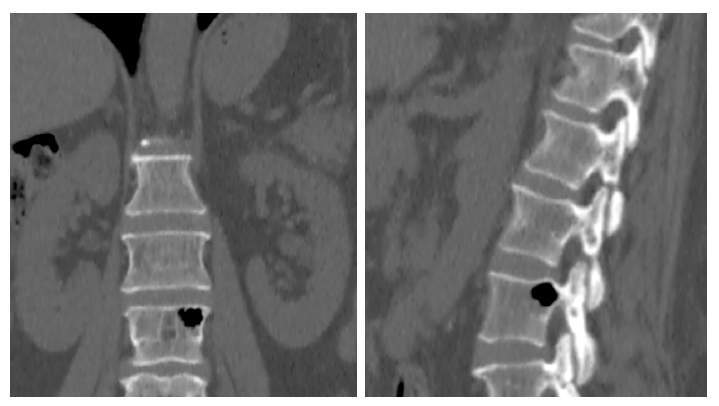
A 57-year old woman requested cancer screening due to family history of liver malignancy. Abdominal computed tomography showed several simple hepatic cysts, and a pneumatized cystic image in the lumbar vertebral body without additional evidence of spinal or intervertebral space injury. Pneumatocysts are usually found in the ileum and the sacrum, as well as in the sacroiliac joints. Pneumatocysts located in vertebral bodies are benign lesions that must be differentiated from other lesions of the spine containing gas, such as osteomyelitis, osteonecrosis, cancer, and post-traumatic and post-surgical causes. The etiology and natural history of pneumatocysts are poorly understood. The diagnosis is based on computed tomography and magnetic resonance images.
