


 Spanish (pdf)
 Article in xml format
 Article references
 Send this article by e-mail
 Cited by SciELO
  Similars in
SciELO
Similars in
SciELO 
 Permalink
Se muestran imagenes tomograficas de dos pacientes de 65 años con diagnóstico confirmado de COVID-19 mediante prueba molecular (Figura 1), ambos con obesidad. El primer paciente presentó un cuadro leve con fiebre, tos y diarrea; el segundo presentó un cuadro severo con fiebre, tos y disnea. En las imagenes tomograficas de tórax en vista axial, sin uso de contraste en ventana pulmonar, se observa el signo de "preservación subpleural" (flechas blancas). Este signo se define como una región de 1-2 mm de parenquima pulmonar subpleural no opacificado, hallazgo muy sugestivo de neumonia intersticial inespecifica. Se observa tambien en casos de contusión pulmonar, proteinosis alveolar pulmonar y algunas infecciones virales. Recientemente se ha descrito en casos de neumonia por la COVID-19 y ha sido considerado como un signo diagnóstico de esta, por lo que resulta importante su identificación en las tomografias de t6rax de este grupo de pacientes.
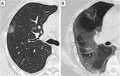
We present tomographic images from two 65-years-old patients with COVID-19 (figure 1), confirmed by molecular testing. Both patients were obese. The first patient developed a mild case with fever, cough and diarrhea; whereas the second patient developed a severe case with fever, cough and dyspnea. The non-enhanced axial tomographic images in lung window show the "subpleural sparing" sign (white arrows). This sign is defined as a 1-2 mm region of non-opacified subpleural pulmonary parenchyma and is highly suggestive of non-specific interstitial pneumonia. It is also observed in pulmonary contusion, pulmonary alveolar proteinosis and some viral infections. Furthermore, it has recently been described in COVID-19 pneumonia and has been considered as a diagnostic sign of this disease, making it important to identify and recognize it on chest tomographic studies from this group of patients.

