SciELO - Scientific Electronic Library Online

 
vol.33 issue2Association between patient satisfaction and motivation of health care personnel in a neurosurgery hospitalization service of a specialized instituteEmotional dependence of students of medicine author indexsubject indexarticles search
Home Pagealphabetic serial listing  

Services on Demand

Journal

Article

Indicators

  • Have no cited articlesCited by SciELO

Related links

  • Have no similar articlesSimilars in SciELO

Share


Revista Medica Herediana

Print version ISSN 1018-130XOn-line version ISSN 1729-214X

Rev Med Hered vol.33 no.2 Lima Apr./Jun. 2022  Epub July 06, 2022

http://dx.doi.org/10.20453/rmh.v33i2.4245 

Comunicación corta

Decompressive craniectomy for malignant middle cerebral artery infarction at an emergency hospital in Peru

Craniectomía descompresiva en infarto maligno de la arteria cerebral media en un hospital de emergencias en Perú

1Departamento de Neurocirugía. Hospital de Emergencias José Casimiro Ulloa, Lima, Perú.

2Departamento de Neurocirugía. Clínica Angloamericana, Lima, Perú.

SUMMARY

Objective

: To describe clinical, surgical and post-operative characteristics of patients with the diagnosis of malignant infarction of the middle cerebral artery who underwent decompressive craniectomy.

Methods

: Descriptive, retrospective case series study, performed between March 2017 and March 2020. Data from consecutive patients with the diagnosis of malignant middle cerebral artery infarction were collected.

Results

: Ten cases were reviewed. Eighty percent of the patients were men, the mean age was 64 years and 60% of the patients were older than 60 years. At admission, the mean Glasgow was 11 points and the mean mRS was 4. The mean time from diagnosis to surgery was 89.7 hours. The anterior cerebral artery was comprised in two cases. Hemorrhagic transformation occurred in three cases. The mean anterior-posterior diameter of the skull flap was 116 mm. The mean ICU and hospital length of stay were 14.1 and 27.5 days, respectively. Three patients died.

Conclusions

: Decompressive craniectomy is a life-saving procedure in an emergency hospital-setting with an acceptable in-hospital mortality rate within one-month follow-up.

KEYWORDS: Middle cerebral artery; decompressive craniectomy; brain ischemia

RESUMEN

Objetivo

: Describir las características clínicas, quirúrgicas y postoperatorias en pacientes con diagnóstico de infarto maligno de la arteria cerebral media sometidos a craniectomía descompresiva.

Material y métodos

: Estudio observacional, descriptivo, retrospectivo, tipo serie de casos, realizado entre marzo 2017 y marzo 2020. Se recolectaron los datos de pacientes consecutivos con diagnóstico de infarto maligno de arteria cerebral.

Resultados

: Diez casos fueron revisados. Ochenta por ciento fueron hombres, la edad promedio fue 64 años y 60% de los pacientes fueron mayores de 60 años. En la admisión, el Glasgow promedio fue de 11 puntos y el mRS fue de 4. El tiempo promedio desde el diagnostico hasta la cirugía fue de 89,7 horas. La arteria cerebral anterior estuvo comprometida en dos casos. La transformación hemorrágica ocurrió en tres casos. El diámetro anteroposterior promedio de la plaqueta ósea fue de 116 mm. El tiempo promedio de estancia en UCI y estancia hospitalaria fueron de 14,1 días y 27,5 días, respectivamente. Tres pacientes murieron.

Conclusiones

: La craniectomía descompresiva es un procedimiento que salva vidas en un ambiente hospitalario de emergencia con una aceptable mortalidad intrahospitalaria dentro del primer mes de seguimiento.

PALABRAS CLAVE: arteria cerebral media; craniectomía descompresiva; isquemia cerebral

INTRODUCTION

Stroke is a leading cause of death and disability worldwide and the fifth cause in the United States of America 1,2. Of all supratentorial cerebrovascular diseases, the malignant infarction of the middle cerebral artery (MCA) can occur in 1 - 10% of the cases. Clinical presentation includes neurological deterioration, progressive neurological deficit, cerebral edema and herniation which can be seen in an ischemic lesion with an arterial territory comprise > 50% with a mortality that can reach 80% 3-7.

Decompressive craniectomy (DC) as part of the treatment for malignant infarction of the MCA continues to be controversial nowadays despite multiple randomized controlled trials showed favorable mortality outcomes but with paradoxical outcomes regarding long-term functional status (modified Rankin score > 3) 8-10.

Therefore, our study aimed to describe clinical, surgical and post-operative characteristics of patients with malignant MCA infarction who underwent DC and assess post-operative outcomes.

METHODS

Observational, descriptive, retrospective study, type case series, carried out between March 2017 and March 2020. Consecutive patients with the diagnosis of malignant stroke of the MCA who underwent DC at the Hospital José Casimiro Ulloa were reviewed. Inclusion criteria included patients with MCA infarction with midline shift and clinical deterioration (Glasgow Coma Scale ( 8).

Demographics and clinical characteristics included age, sex, past medical history, Glasgow and modified Rankin Scale at admission, the presence of anisocoria, motor deficit, blood pressure at admission and time to surgery since diagnosis were evaluated.

The diagnosis of MCA infarction was made based on brain CT images: a hypodensity in the MCA territory with midline shift was assessed by the radiologist, neurologist or neurosurgeon. MRI and CT perfusion were not employed in our institution. In cases of hemorrhagic transformation, the presence of blood within the hypodensity and clinical presentation were assessed. The surgical decision was made by clinical deterioration and if the territory of the MCA infarction was ( 50%.

DC was done based on previous technical principles. A question mark incision was made in the affected side, the skull flap was removed, the dura was opened and then duroplasty was performed. The bone flap was kept in the abdominal subcutaneous tissue. Time of surgery, blood loss and mortality were analyzed.

The study was exempted from the institutional board review due to the retrospective design and review of clinical charts.

RESULTS

Patient characteristics

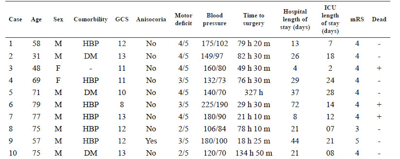
Ten patients were retrospectively reviewed. Eight patients were men. The mean age was 64 ± 15.4 years (range 31 to 79 years); six cases were older than 60 years. The mean Glasgow Coma Scale at the moment of the diagnosis was 11 points (range 8 to 13 points). Anisocoria and motor deficit were present in one and ten patients, respectively. The mean systolic pressure was 156.7 mmHg and the mean diastolic pressure was 95.6 mmHg at the time of the diagnosis. The mean time from diagnosis of stroke to surgery was 89.7 hours. The mean preoperative modified Rankin score was 4. All the patients had right dominance. Overall characteristics of the patients are shown in Table 1.

Table 1 Clinical characteristics of patients who underwent decompressive craniectomy for malignant middle cerebral artery infarction. 

HBP: high blood pressure, DM: diabetes mellitus

GCS: Glasgow Coma Scale

ICU: Intensive Care Unit

mRS: modified Rankin Scale

Stroke characteristics

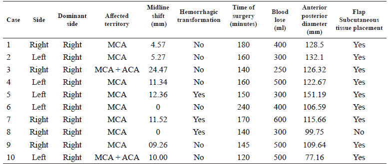
The left middle cerebral artery was affected in 50% of the cases. In all cases the MCA was affected and in two cases the anterior cerebral artery was also comprised. Midline shift was present in eight cases with a mean shift of 8.9 mm. Three patients had a hemorrhagic transformation of the stroke. An illustrative case is shown in figure 1. Radiological characteristics are shown in table 2.

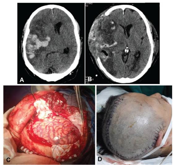
Figure 1 A: Brain CT scan shows a malignant infarction of the right middle cerebral artery with hemorrhagic transformation. B: Brain CT scan following decompressive craniectomy 72 hours after hospital admission. C-D: Intraoperative dura opening and post-operative skin incision. 

Table 2 Radiological and surgical characteristics of patient who underwent decompressive craniectomy for malignant middle cerebral artery infarction. 

Surgical characteristics, post-operative evolution and complications

DC was done in the left side in 50% of the cases. The mean anterior-posterior diameter of the bone flap was 116 mm ( 20.3 mm (range 77.2 mm to 151.2 mm). The average time of surgery was 151.5 minutes and the mean blood loss was 405 ml. In nine patients the skull flap was kept in the abdominal subcutaneous tissue. The mean ICU length of stay was 14.1 days (range 2 to 28 days). The mean hospital length of stay was 27.5 days (range 4 to 72 days). No patient improved clinically at discharge. Three patients died (30%), of which two of them were older than 60 years.

DISCUSSION

According to our results, the in-hospital mortality rate after performing DC in patients with malignant MCA infarction was 30% and time to surgery since the initial diagnosis of stroke was 89.7 hours. DC is a life-saving neurosurgical procedure used in cases of increased intracranial pressure due malignant infarction of the MCA. Previous papers have shown the benefits of this procedure in mortality but not in functional recovery.

Three controlled randomized trials have evaluated the rationale of DC in cases of MCA strokes. The DESTINY trial evaluated 32 patients and determined a considerable mortality reduction in their analysis (88% of non-DC versus 47% of patients who underwent DC 8, the DECIMAL trial evaluated 38 patients with a survival rate favoring patients who underwent DC (75% versus 22%) 9, and the HAMLET trial evaluated 64 patients in which an ARR of 38% in mortality favored the DC-group 10.

Overall, these studies found that patients under 60 years had a lower mortality rate at 12 months when compared with medical management. In addition, Vahedi et al. 11, performed a pooled analysis of these three RCT and determined a considerable reduction in mortality (22% versus 71%), no improvement in the proportion of survivors with good outcomes and no difference in clinical outcomes among dominant and non-dominant hemispheres. On the other hand, DESTINY II trial evaluated patients older than 60 years and their results showed that survival without severe disability was higher in the DC-group in comparison to the control group (38% versus 18%). Although, overall mortality was lower in the DC-group (33% versus 70%) 12 .Other studies also shown poor outcomes in patients older than 60 years 13. Nevertheless, other authors reported better functional outcomes and lower mortality rates in this subgroup of patients 14,15.

In addition, the variable time to surgery since the initial diagnosis of stoke does not reflect the time elapsed from malignant stroke development to surgery.

In our country, few studies have evaluated in-hospital mortality rates due to cerebrovascular diseases. In a national hospital, the overall mortality rate was 19.6%, most of them within the first three days 16, whereas another study reported a mortality of 20.8% 17. Obviously, these studies did not evaluate mortality in patients with MCA occlusion who underwent DC. Conversely, they report a general overview of cerebrovascular diseases and set a landmark for future investigations in the cerebrovascular field. Despite these results, a recent paper reported a decreased tendency which was heterogeneous among locations within the same country 18. Previous studies reported that patients who underwent DC with larger bone flaps showed better outcomes 19-21. This is particularly special in traumatic brain injury cases 22,23. We found that our mean anterior-posterior diameter of the bone flap was 116 mm. Two patients who died had a skull flap less than 12 cm.

The study has several limitations: the small number of patients, the retrospective design and the lack of longer clinical follow-up are the most important. Our center is an emergency hospital where life-saving surgeries are performed and patients are therefore transferred to other centers or discharged home when they recover, making mid- and long-term clinical follow-up unviable.

DC is a life-saving procedure performed in an emergency-setting with an adequate in-hospital mortality rate despite time to diagnosis and age. Mid-term and long-term follow-ups are needed in this setting. Future directions should be established in order to improve the public services in developing countries in terms of neurosurgical care.

REFERENCES

1. Johnson CO, Nguyen M, Roth GA, et al. Global, regional, and national burden of stroke, 1990-2016: a systematic analysis for the Global Burden of Disease Study 2016. Lancet Neurol. 2019; 18(5):439-58. [ Links ]

2. Benjamin E, Muntner P, Alonso A, et al. Heart Disease and Stroke Statistics-2019 Update: A Report From the American Heart Association. Circulation. 2019; 139(10): e56-528. [ Links ]

3. Hacke W, Schwab S, Horn M, Spranger M, De Georgia M, von Kummer R. "Malignant" Middle Cerebral Artery Territory Infarction: Clinical Course and Prognostic Signs. Arch Neurol. 1996; 53(4):309-15. [ Links ]

4. Treadwell SD, Thanvi B. Malignant middle cerebral artery (MCA) infarction: pathophysiology, diagnosis and management. Postgrad Med J. 2010; 86(1014): 235 LP-242. [ Links ]

5. Huttner HB, Schwab S. Malignant middle cerebral artery infarction: clinical characteristics, treatment strategies, and future perspectives. Lancet Neurol. 2009;8(10):949-58. [ Links ]

6. Wartenberg KE. Malignant middle cerebral artery infarction. Curr Opin Crit Care. 2012; 18(2) :152-63. doi: 10.1097/MCC.0b013e32835075c5 [ Links ]

7. Kasner S, Demchuk, Berrouschot J, et al. predictors of fatal brain edema in massive hemispheric ischemic stroke. Stroke. 2001; 32(9):2117-23. [ Links ]

8. Juttler E, Schwab S, Schmiedek P, et al. Decompressive Surgery for the Treatment of Malignant Infarction of the Middle Cerebral Artery (DESTINY): a randomized, controlled trial. Stroke. 2007; 38(9):2518-25. [ Links ]

9. Vahedi K, Vicaut E, Mateo J, et al. sequential-design, multicenter, randomized, controlled trial of early decompressive craniectomy in malignant middle cerebral artery infarction (DECIMAL Trial). Stroke. 2007; 38(9):2506-17. [ Links ]

10. Hofmeijer J, Kappelle LJ, Algra A, Amelink GJ, van Gijn J, van der Worp HB. Surgical decompression for space-occupying cerebral infarction (the Hemicraniectomy After Middle Cerebral Artery infarction with Life-threatening Edema Trial [HAMLET]): a multicentre, open, randomised trial. Lancet Neurol. 2009; 8(4):326-33. [ Links ]

11. Vahedi K, Hofmeijer J, Juttler E, et al. Early decompressive surgery in malignant infarction of the middle cerebral artery: a pooled analysis of three randomised controlled trials. Lancet Neurol. 2007; 6(3):215-22. [ Links ]

12. Jüttler E, Unterberg A, Woitzik J, et al. Hemicraniectomy in older patients with extensive middle-cerebral-artery stroke. N Engl J Med. 2014;3 70(12):1091-100. [ Links ]

13. Arac A, Blanchard V, Lee M, Steinberg GK. Assessment of outcome following decompressive craniectomy for malignant middle cerebral artery infarction in patients older than 60 years of age. Neurosurg Focus FOC. 2009; 26(6):E3. [ Links ]

14. Yu JW, Choi J-H, Kim D-H, Cha J-K, Huh J-T. Outcome following decompressive craniectomy for malignant middle cerebral artery infarction in patients older than 70 years old. J Cerebrovasc Endovasc Neurosurg. 2012; 14(2):65-74. [ Links ]

15. Zhao J, Su YY, Zhang Y, et al. Decompressive hemicraniectomy in malignant middle cerebral artery infarct: a randomized controlled trial enrolling patients up to 80 years old. Neurocrit Care. 2012; 17(2):161-71. [ Links ]

16. Castañeda-Guarderas A, Beltrán-Ale G, Casma-Bustamante R, Ruíz-Grosso P, Málaga G. Registro de pacientes con accidente cerebro vascular en un hospital público del Perú, 2000-2009. Rev Peru Med Exp Salud Publica. 2011; 28(4):623-7. [ Links ]

17. Alvarado-Dulanto CMA, Lazo M, Loza-Herrera JD, Malaga G. Pronóstico al año tras sufrir el debut de enfermedad cerebrovascular en pacientes de un hospital nacional de Lima, Perú. Rev Peru Med Exp y Salud Pública; 2015; 32(1):98-103. [ Links ]

18. Atamari-Anahui N, Alva-Diaz C, Vera-Monge V, Taype-Rondan A. Tendencia de mortalidad por enfermedad cerebrovascular registrada por el Ministerio de Salud de Perú, 2005-2015. Neurol Argentina. 2019; 11(4):202-9. [ Links ]

19. Neugebauer H, Fiss I, Pinczolits A, et al. Large size hemicraniectomy reduces early herniation in malignant middle cerebral artery infarction. Cerebrovasc Dis. 2016; 41(5-6):283-90. [ Links ]

20. Chung J, Bang OY, Lim YC, Park SK, Shin YS. Newly suggested surgical method of decompressive craniectomy for patients with middle cerebral artery infarction. Neurologist. 2011; 17(1):11-15. [ Links ]

21. Delashaw JB, Broaddus WC, Kassell NF, et al. Treatment of right hemispheric cerebral infarction by hemicraniectomy. Stroke. 1990; 21(6):874-81. [ Links ]

22. Sedney CL, Julien T, Manon J, Wilson A. The effect of craniectomy size on mortality, outcome, and complications after decompressive craniectomy at a rural trauma center. J Neurosci Rural Pract. 2014; 5(3):212-7. [ Links ]

23. Huang X, Wen L. Technical considerations in decompressive craniectomy in the treatment of traumatic brain injury. Int J Med Sci. 2010; 7(6):385-90. [ Links ]

Received: October 31, 2020; Accepted: January 13, 2022

Correspondencia: Giancarlo Saal-Zapata Avenida Roosevelt 6355, Miraflores, Lima, Perú. e-mail: gian_carlo1987@hotmail.com

Declaración de financiamiento y conflictos de interés:

Reporte fue autofinanciado por los autores. No existe conflicto de intereses de parte de los autores ni de orden personal, económico, institucional o laboral.

Contribución de autoría:

GSZ: Concepción y redacción del artículo, recolección e interpretación de los datos, cirujano participante del procedimiento y aprobación de la versión final. FC: Concepción y redacción del artículo, recolección e interpretación de los datos, cirujano participante del procedimiento y aprobación de la versión final.

Creative Commons License This is an open-access article distributed under the terms of the Creative Commons Attribution License