SciELO - Scientific Electronic Library Online

 
vol.30 número4La tradición de calidad educativa del Programa de EstomatologíaEmpleabilidad de los egresados de Estomatología índice de autoresíndice de materiabúsqueda de artículos
Home Pagelista alfabética de revistas  

Servicios Personalizados

Revista

Articulo

Indicadores

  • No hay articulos citadosCitado por SciELO

Links relacionados

  • No hay articulos similaresSimilares en SciELO

Compartir


Revista Estomatológica Herediana

versión impresa ISSN 1019-4355

Rev. Estomatol. Herediana vol.30 no.4 Lima oct-dic 2020

http://dx.doi.org/10.20453/reh.v30i4.3877 

Original articles

Strategies and operational procedures in oral radiology to mitigate potential SARS-CoV-2 transmission

Estrategias y procedimientos operacionales en Radiología Oral para mitigar transmisión de SARS-CoV-2

Gina Delia Roque-Torres1  , DDS, MSc, PhD. Assistant Professor

Victor Toral Rizo2  , DDS, MSc, PhD

Constanza E. Fernández3  , DDS, MSc, PhD

Cristian Camilo Madrid Troconis4 

Eder Alberto Sigua-Rodriguez5  , DDS, MSc, PhD

Stephanie Perez Puello6 

Gustavo Tello Melendez7  , DDS, MSc, PhD

1 Center of Dental Research, School of Dentistry, Loma Linda University. Loma Linda, California, United States of America.

2 Center for Research and Advanced Studies in Dentistry, School of Dentistry, Autonomous University of the State of Mexico, Toluca, State of Mexico, Mexico.

3 Cariology Unit, Department of Oral Rehabilitation, School of Dentistry, Faculty of Oral Health, University of Talca, Talca, Chile.

4 University of Cartagena, Department of Oral Rehabilitation, School of Dentistry, Cartagena, Colombia.

5 Research Department, Institución Universitaria Colegios de Colombia Bogotá, Colombia.

6 Corporación Universitaria Rafael Nuñez, Social and Community Dentistry Department, School of Health Sciences,Cartagena, Colombia.

7 Paediatric Dentistry Department, University of São Paulo. São Paulo, Brazil.

SUMMARY

Coronavirus disease (COVID-19) is highly transmissible through respiratory droplets, saliva, and aerosol particles. Infection can lead to severe acute respiratory syndrome and trigger multi-organ failure and death. Most clinical dental procedures involve contact with saliva or the generation of aerosols with saliva and blood. These airborne particles increase the risk of transmission by COVID-19 between dentists and patients. Thus, dentistry must adopt procedures to reduce transmission during dental procedures. Although these strategies may not be directly applicable to all institutions due to cultural, social, geographical, and economic differences, the underlying principles and implemented strategies remain relevant and can be adopted or modified by the institutions dealing with the current COVID-19 outbreak. The purpose of this article is to discuss one of the principal sources of COVID-19 transmission: saliva, the biofluid most frequently handled in a radiology environment. The authors also recommend strategies and operational procedures to decrease the spread of COVID-19 in a radiological routine.

KEYWORDS: Diagnosis; detection; betacoronavirus; saliva; coronavirus infections; infection control; radiology

RESUMEN

La enfermedad por coronavirus (COVID-19) se transmite fácilmente a través de gotitas respiratorias, saliva y partículas de aerosol. La infección puede desencadenar un síndrome respiratorio agudo severo, insuficiencia multiorgánica y, en algunos casos, la muerte. Los procedimientos clínicos dentales determinan el contacto con saliva o la generación de aerosoles con saliva y sangre, lo que implica un alto riesgo de transmisión por COVID-19 entre odontólogos y pacientes. Por ello, la práctica odontologica debe adoptar medidas para detener la propagación durante los procedimientos dentales. En este artículo se revisan las estrategias para mitigar el riesgo de transmisión de COVID-19. Si bien estas estrategias pueden no ser directamente aplicables a todas las instituciones debido a diferencias culturales, sociales, geográficas y económicas inherentes, los principios subyacentes y las estrategias implementadas siguen siendo relevantes y pueden ser adoptados o modificados por las instituciones que se ocupan del brote actual de COVID-19. El propósito de este artículo es discutir una de las principales fuentes de transmisión de COVID-19: la saliva, el biofluido que se maneja con mayor frecuencia en un entorno radiológico. Los autores también recomiendan estrategias y procedimientos operativos para disminuir la propagación de COVID-19 en una rutina radiológica.

PALABRAS CLAVE: Diagnóstico; detección betacoronavirus; saliva; infecciones por coronavirus; control de infecciones; radiología.

Epidemiologic Characteristics and Transmission

In December 2019, a group of individuals diagnosed with pneumonia without etiology were reported in Wuhan, China, and later labeled as severe acute respiratory syndrome coronavirus 2 (SARS-Cov-2) (1-3). The original symptoms of SARS-Cov-2 infection included fever, cough, and shortness of breath within 2 to 14 days after exposure 4. Other symptoms have been added later including such as fatigue, muscle or body aches, headache, loss of taste or smell, chest pain, sore throat, congestion or runny nose, nausea or vomiting, diarrhea and a rash on the skin, or discoloration of fingers or toes. 5

In the following weeks the infection spread to other Asian countries, Europe, North America, South America, and Africa, for which, on March 11, 2020, the World Health Organization (WHO) declared the disease COVID-19 a pandemic 5. As of December 2020; 68,812,210 confirmed cases of COVID-19 were detected worldwide, and 1,567,297 deaths have been reported 6.

The virus is highly contagious and spreads person-to-person through close contact, respiratory droplets, and aerosol particles.

Coronaviruses are part of the Coronaviridae family in the Nidovirales order 7. The outer surface of the virus contains spike-like proteins resembling a crown (corona). These proteins facilitate the link with receptors in the respiratory epithelial cells. In size, SARS-CoV-2 is small (65-125 nm in diameter), holds a single-stranded RNA of 26 to 32 kbs. There are four subgroups of the coronavirus family: alpha (α), beta (β), gamma (γ) and delta (ᵟ) 8.

The transmission of SARS-Cov-2 occurs from asymptomatic, presymptomatic, or symptomatic infected individuals 9. Person-to-person spreading occurs due to close contact with an infected person who is coughing, sneezing, or spreading respiratory droplets, and aerosols. These aerosols can enter the human body via inhalation through the nose or mouth and then go to the lungs 8,10. The angiotensin-converting enzyme 2 (ACE2), in the cells of the lower respiratory tract of humans, acts as a cell receptor for SARS-CoV-2 11,12. Wan et al., identified this receptor-like regulator in a cross-species human-to-human transmission 13. This ACE2 receptor has been identified also in the human oral mucosa cells (14-16), thus the oral cavity could be regarded as susceptible to SARS-CoV-2 infection 14,17,18. The incubation period after infection is 1-14 days, but 3-7 days in most cases. During the incubation time, the infected person is infectious 19,20. The virus has been detected in saliva 28.

Diagnosis and Detection of COVID-19

The clinical and public health needs of the pandemic required rapid and reliable tests to detect persons infected with SARS-CoV-2 29. At present, real-time reverse transcription-polymerase chain reaction (rRT-PCR) on respiratory specimens represents the reference standard test for the detection of SARS-CoV2 infection 27. To perform rRT-PCR, the sample is obtained through a nasopharyngeal swab 17. Sputum and oropharyngeal secretions have been suggested as possible targets for the molecular diagnosis of COVID-19 by rRT-PCR 28. Nasopharyngeal swabs require close contact between tested individuals and healthcare workers who may be at an increased risk of exposure if not donning appropriate barriers. Moreover, these types of specimens may also cause discomfort, and even bleeding in patients with low levels of thrombocytes 27,28.

Saliva has been used as a diagnostic tool for a variety of diseases 24,-32), conditions 36-38, and viral infections 33-35. Some studies have validated saliva as a tool to detect SARS-CoV-2 against (insert the comparison) 16,22,28,39 and reported sensitivity and specificity of X%, and Y%, respectively 28.

As a source of SARCS-CoV-2, saliva has some additional advantages: it remains stable at room temperature for up to 24 hours and up to a week at 4 °C 21. Furthermore, obtaining a saliva sample does not cause discomfort to patients, making it ideal for pediatric patients 22,40, by not requiring nasal swabs 24.

SARS-Cov-2 Transmission to Dental Practitioners

Dental practitioners are at higher risk of infection by SARS-CoV-2 because most dental procedures produce contaminated splatters, droplets, and aerosols 25. This is particularly important when providing care to asymptomatic or presymptomatic patients 17,27. Intraoral imaging is the most common radiographic technique; thus, care should be taken not to stimulate saliva secretion or coughing 54. In their interim recommendations to reduce transmission in dental settings. the U.S. Centers for Disease Control and Prevention suggest the use of extraoral instead of intraoral imaging, along with appropriate personal protection equipment (PPE). However, panoramic radiography and cone-beam computed tomography (CBCT) may expose the individual to higher levels of radiation than a single periapical image 56 or may not be a suitable alternative for intraoral imaging.

Several publications have reported on the radiological characteristics of COVID-19 individuals 41-45, and the recommendations on how to take their images. 46-52. Only one publication has provided specific recommendations for dental radiology 53.

Proposed Appointment Model for Oral-Dental Radiology

In the next section, we provide general guidance on how to devise a radiology appointment. The general recommendation of keeping social distance during the time the patient is in pre-clinical and clinical area as well as wearing mask applies. These recommendations, should be in concordance with institutional and national policies, 57 and communicated to the patient before the appointment 58.

Preappointment (Figure 1)

Figure 1 Prescan workflow 

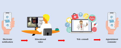
1. Electronic notification of appointment date and time.

2. If possible, share with the patient informational videos on the radiographic procedures to be used.

3. Tele-consult to screen for symptoms, risk behaviors and potential exposures to COVID-19 follow institutional recommendations on what to ask and how, like the ones CDC recommended:

a. Have you experienced any of the following symptoms in the past 48 hours: (fever or chills, cough, shortness of breath or difficulty breathing, fatigue, muscle or body aches, headache, new loss of taste or smell, sore throat, congestion or runny nose, nausea or vomiting, diarrhea)

b. Within the past 14 days, have you been in close physical contact (6 feet or closer for a cumulative total of 15 minutes) with: Anyone who is known to have laboratory-confirmed COVID-19? OR Anyone who has any symptoms consistent with COVID-19?

c. Are you isolating or quarantining because you may have been exposed to a person with COVID-19 or are worried that you may be sick with COVID-19?

d. Are you currently waiting on the results of a COVID-19 test?

4. Send a reminder close to the appointment date with information regarding wearing masks and what to do upon arrival. Is common practice by most dental clinics to space the appointments so reduce crowding in the waiting area. Others require the patient to wait outside the office.

5. Scan and go concept: This concept aims to complete all administrative affairs before the clinical procedure 48. Thus, transactions such as payments or others should be handled electronically.

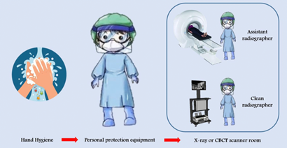
During the appointment (Figure 2)

Figure 2 During the scanning workflow. 

Health questionnaire and history of potential risks after the teleconsultation. For this step, the same questions from the pre-consult could be used.

Measuring body temperature.

The patient is led to the radiological unit.

Patients and any companion should be wearing masks while in the office and receive appropriate standard radiation protection devices 59. Other recommendations for protecting surfaces in the office apply 60.

5. Use two radiographers: a radiographer assigned to operate the workstation and controls, and an assistant radiographer assigned to interact with the patient during clinical procedures 46,61,62.

6. Patients can mouthwash with very diluted hydrogen peroxide (0.75%) or long-lasting iodine solutions before the procedure, but there is no supporting evidence that these procedures reduce transmission of COVID-19 63.

7. There is no need to remove the mask during extraoral scans.

Post appointment

1. Disinfection of all surfaces: COVID-19 can persist on surfaces for hours or days, depending on the type of surface, temperature, or humidity of the environment 64. Thus, all the essential items in the room i.e. operating consoles such as monitors, keyboards, mouse, chairs, control panel, and exposure buttons or sensors should be covered with waterproof protective material. At the end of the radiological procedure, all covering material should be removed and disposed. Areas that have not been covered must be reprocessed with standard hospital disinfectant: low- or intermediate-level disinfectants, such as iodophor germicidal detergent solution, ethyl alcohol 70% or 75%, or isopropyl alcohol, following the equipment manufacturers' recommendations 46,49,65. Removing non-essential items from the clinical area reduce reprocessing time.

2. The scan room needs to be closed tightly sealed for 15 min to thoroughly ventilate and exchange the room air 60.

3. If necessary, consultation with the patient, family, colleagues should be done by phone or e-consult platforms 50.

4. Practitioners should review periodically the updates on epidemiological, clinical, and infection control information from reputable sources websites.

5. Reporting results from the radiological examination could be done electronically by radiology information system (RIS), or picture archiving and communication system (PACS), as well as an update to the patient via the online portal 46. The practitioner should be aware of legal requirements for sharing personal and clinical information and secure appropriate sign consent from the patient.

All the above is in concurrence with the recommended routine infection prevention and control practices during the COVID-19 pandemic released by the Center for Disease Control and Prevention (CDC) and the World Health Organization (WHO), such as implementing telehealth, screening triage, implement universal source control measures, and physical distance at least 6 feet between people, and performing only elective procedures during increased transmission in the population. 59,60,66

Impacts of COVID-19 on the oral radiology specialty

The original restrictions on clinical practice due to the pandemic affected the oral radiology specialty by reducing encounters to those required under emerging services only. In the medical field, experiences from other countries suggest a 50% to 70% decrease in imaging volume that lasts a minimum of 3-4 months 67. It is still too early to clarify the economic impact on dental radiology practices and services, especially when in most countries dental care has returned close to pre-pandemic figures. The American Dental Association (ADA) predicts that U.S. dental care spending could decline by up to 38 percent in 2020 and 20 percent in 2021 68. The most recent ADA data are encouraging since the modeling predicts that U.S. dental care spending could decline by up to 38 percent in 2020 and 20 percent in 2021. However, it could be expected that dental practices may be affected if additional restrictions are set in response to new outbreaks.

COVID-19 pandemic and its affect on clinical research

There are no reliable reports on the effect of COVID-19 on oral and craniofacial research. However, anecdotal information suggests that most clinical trials were postponed, or have interrupted their protocols in their inability to take direct measurements on participants in research centers or dental schools, due to lockdowns. Researchers have opted for conducting other aspects of their protocols to save time, 46,52,70. but intraoral interventions or measurements were postponed due to the pandemic, including those requiring imaging. Some have predicted loss of scientific productivity for as much as 1 - 2 years as the result of a shutdown 53,70. The effect will be particularly acute in clinical trials, where investigators will deal with missed visits, the need to re-engage existing subjects, having to recruit new subjects, and protocol alterations as reported in the medical field 70.

In the field of radiology, COVID-19-related research has increased to create components for personal protection equipment (PPE) and other devices 70. The need from several countries and healthcare institutions for these alternatives and the subsequent lack of supply might be addressed by the use of three-dimensional (3D) printing. It has become the solution to the shortage of PPE 71, even if none studies are describing the effectiveness.

Final Comments and Conclusions

Further studies are necessary to ascertain the health and economic impact of limiting dental care during COVID-19 restrictions, including oro- and maxilla-facial radiology. The magnitude of the pandemic has stretched the limited available resources and open the field for novel technologies.

Uncertainty as a consequence of the COVID-19 pandemic is demanding the profession to innovate. The most recent data from the U.S. suggest that practices are returning to levels of pre-pandemic busyness. However, new outbreaks will be the norm in the future. If there is anything the profession has learned from this pandemic, is that we were not prepared. It is necessary to invent, adapt, or improve new equipment, protective barriers, and disinfectants that will allow the delivery of dental care under safety standards. Ultimately, it will be the result of cooperation and agreement between the government, dental associations, academic institutions, and individual practitioners which will determine new practice environments, alternative clinical procedures in what we can call the new dentistry in 2021. In this document, we propose some of the procedures for the specialty of oral radiology.

REFERENCES

1. World Health Organization. Pneumonia of unknown cause - China. Geneva: World Health Organization; 2020. (Cited 2020 May 20). Available from: https://www.who.int/csr/don/05-january-2020-pneumonia-of-unkown-cause-china/en/. [ Links ]

2. McMichael TM, Currie DW, Clark S, Pogosjans S, Kay M, Schwartz NG, et al. Epidemiology of Covid-19 in a Long-Term Care Facility in King County, Washington. N Engl J Med. 2020;1: 1-7. [ Links ]

3. Zhu N, Zhang D, Wang W, et al. A novel coronavirus from patients with pneumonia in China, 2019. N Engl J Med. 2020;382(8):727-33. [ Links ]

4. Centers for Disease Control and Prevention. Coronavirus Disease 2019 in Children. Morbidity and Mortality Weekly Report (MMWR). 2020;66:2019-21. (Cited 2020 May 20). Available from: https://www.cdc.gov/mmwr/volumes/69/wr/mm6914e4.htmLinks ]

5. World Health Organization. Coronavirus (COVID-19) events as they happen. Geneva: World Health Organization; 2020. (cited 2020 May 19). Available from: https://www.who.int/emergencies/diseases/novel-coronavirus-2019/events-as-they-happen [ Links ]

6. Johns Hopkins Coronavirus Resource Center. COVID-19 Map. Baltimore: Johns Hopkins Coronavirus Resource Center; 2020. (Cited 2020 May 20). Available from: https://coronavirus.jhu.edu/map.html [ Links ]

7. Weston S, Frieman MB. Respiratory Viruses. In: Encyclopedia of Microbiology. New York: Elsevier; 2019. (Cited 2020 May 20). Available from: https://linkinghub.elsevier.com/retrieve/pii/B9780128012383661615 [ Links ]

8. Shereen MA, Khan S, Kazmi A, Bashir N, Siddique R. COVID-19 infection: Origin, transmission, and characteristics of human coronaviruses. J Adv Res. 2020;24:91-8. Doi: 10.1016/j.jare.2020.03.005 [ Links ]

9. Bai Y, Yao L, Wei T, et al. Presumed asymptomatic carrier transmission of COVID-19. JAMA. 2020;323(14):1406-1407. [ Links ]

10. Parry J. China coronavirus: cases surge as official admits human to human transmission. BMJ. 2020;368:m236. [ Links ]

11. Jia HP, Look DC, Shi L, et al. ACE2 Receptor Expression and Severe Acute Respiratory Syndrome Coronavirus Infection Depend on Differentiation of Human Airway Epithelia. J Virol. 2005;79(23):14614-21. doi: 10.1128/JVI.79.23.14614-14621.2005 [ Links ]

12. Zhou P, Yang X Lou, Wang XG, et al. A pneumonia outbreak associated with a new coronavirus of probable bat origin. Nature. 2020;579(7798):270-3. Doi: 10.1038/s41586-020-2012-7. [ Links ]

13. Wan Y, Shang J, Graham R, Baric RS, Li F. Receptor Recognition by the Novel Coronavirus from Wuhan: an Analysis Based on Decade-Long Structural Studies of SARS Coronavirus. J Virol. 2020;94(7): e00127-20. doi: 10.1128/JVI.00127-20 [ Links ]

14. Xu H, Zhong L, Deng J, et al. High expression of ACE2 receptor of 2019-nCoV on the epithelial cells of oral mucosa. Int J Oral Sci. 2020;12(1):8. Doi: 10.1038/s41368-020-0074-x [ Links ]

15. Zhang H, Kang Z, Gong H, et al. The digestive system is a potential route of 2019-nCov infection: a bioinformatics analysis based on single-cell transcriptomes. bioRxiv. 2020. Doi: 10.1136/gutjnl-2020-320953 [ Links ]

16. Zhao Y, Zhao Z, Wang Y, Zhou Y, Ma Y, Zuo W. Single-cell RNA expression profiling of ACE2, the putative receptor of Wuhan 2019-nCov. bioRxiv. 2020;2020.01.26.919985. doi: 10.1101/2020.01.26.919985 [ Links ]

17. Azzi L, Carcano G, Gianfagna F, et al. Saliva is a reliable tool to detect SARS-CoV-2. J Infect. 2020;81(1):e45-e50. doi: 10.1016/j.jinf.2020.04.005 [ Links ]

18. Xu J, Li Y, Gan F, Du Y, Yao Y. Salivary Glands: Potential Reservoirs for COVID-19 Asymptomatic Infection. J Dent Res. 2020; 99(8):989. doi: 10.1177/0022034520918518 [ Links ]

19. Guo Y-R, Cao Q-D, Hong Z-S, et al. The origin, transmission and clinical therapies on coronavirus disease 2019 (COVID-19) outbreak - an update on the status. Mil Med Res. 2020;7(1):11. doi: 10.1186/s40779-020-00240-0 [ Links ]

20. Jin YH, Cai L, Cheng ZS, , et al. A rapid advice guideline for the diagnosis and treatment of 2019 novel coronavirus (2019-nCoV) infected pneumonia (standard version). Med J Chinese People's Lib Army. 2020;45(1):1-20. [ Links ]

21. Kaufman E, Lamster IB. The diagnostic applications of saliva--a review. Crit Rev Oral Biol Med. 2002;13(2):197-212. doi: 10.1177/154411130201300209. [ Links ]

22. Khurshid Z, Asiri FYI, Al Wadaani H. Human Saliva: Non-Invasive Fluid for Detecting Novel Coronavirus (2019-nCoV). Int J Environ Res Public Health. 2020;17(7):2225. doi: 10.3390/ijerph17072225 [ Links ]

23. Martina E, Campanati A, Diotallevi F, Offidani A. Saliva and Oral Diseases. J Clin Med. 2020;9(2):466. [ Links ]

24. Chojnowska S, Baran T, Wilinska I, Sienicka P, Cabaj-Wiater I, Knas M. Human saliva as a diagnostic material. Adv Med Sci. 2018;63(1):185-191. doi: 10.1016/j.advms.2017.11.002 [ Links ]

25. Li Y, Ren B, Peng X, et al. Saliva is a non-negligible factor in the spread of COVID-19. Mol Oral Microbiol. 2020;35(4):141-145. doi: 10.1111/omi.12289 [ Links ]

26. Fernandes LL, Pacheco VB, Borges L, et al. Saliva in the Diagnosis of COVID-19: A Review and New Research Directions. J Dent Res. 2020;22034520960070. doi: 10.1177/0022034520960070. Online ahead of print. [ Links ]

27. Wang Y, Kang H, Liu X, Tong Z. Combination of RT-qPCR testing and clinical features for diagnosis of COVID-19 facilitates management of SARS-CoV-2 outbreak. J Med Virol. 2020;2:538-9. [ Links ]

28. To KKW, Tsang OTY, Chik-Yan Yip C, et al. Consistent detection of 2019 novel coronavirus in saliva. Clin Infect Dis. 2020;1:4-6. doi: 10.1093/cid/ciaa149 [ Links ]

29. Chan JFW, Yuan S, Kok KH, et al. A familial cluster of pneumonia associated with the 2019 novel coronavirus indicating person-to-person transmission: a study of a family cluster. Lancet. 2020;395(10223):514-23. Doi: 10.1016/S0140-6736(20)30154-9 [ Links ]

30. Nunes LAS, Mussavira S, Bindhu OS. Clinical and diagnostic utility of saliva as a non-invasive diagnostic fluid: A systematic review. Biochem Medica. 2015;25(2):177-92. [ Links ]

31. Zhang L, Xiao H, Karlan S, et al. Discovery and preclinical validation of salivary transcriptomic and proteomic biomarkers for the non-invasive detection of breast cancer. PLoS One. 2010;5(12):1-7. [ Links ]

32. Xiao H, Zhang L, Zhou H, Lee JM, Garon EB, Wong DTW. Proteomic analysis of human saliva from lung cancer patients using two-dimensional difference gel electrophoresis and mass spectrometry. Mol Cell Proteomics. 2012;11(2):1-12. [ Links ]

33. Corstjens PLAM, Abrams WR, Malamud D. Detecting viruses by using salivary diagnostics. J Am Dent Assoc. 2012;143(10 Suppl):12S-8S. doi: 10.14219/jada.archive.2012.0338 [ Links ]

34. Balamane M, Winters MA, Dalai SC, et al. Detection of HIV-1 in Saliva: Implications for Case-Identification, Clinical Monitoring and Surveillance for Drug Resistance. Open Virol J. 2010;4:88-93. doi: 10.2174/1874357901004010088 [ Links ]

35. Khadse SV, Bajaj G, Vibhakar P, Nainani P, Ahuja R, Deep G. Evaluation of specificity and sensitivity of oral fluid for diagnosis of hepatitis B. J Clin Diagnostic Res. 2016;10(1):BC12-4. [ Links ]

36. Tao D, McGill B, Hamerly T, et al. A saliva-based rapid test to quantify the infectious subclinical malaria parasite reservoir. Sci Transl Med. 2019;11(473). [ Links ]

37. Sreebny L, Zhu WX. Whole saliva and the diagnosis of Sjögren's syndrome: an evaluation of patients who complain of dry mouth and dry eyes. Part 1: Screening tests. Gerodontology. 1996;13(1):35-43. [ Links ]

38. Bonamico M, Nenna R, Montuori M, et al. First salivary screening of celiac disease by detection of anti-transglutaminase autoantibody radioimmunoassay in 5000 Italian primary schoolchildren. J Pediatr Gastroenterol Nutr. 2011;52(1):17-20. [ Links ]

39. Vinayachandran D, Balasubramanian S. Salivary diagnostics in COVID-19: Future research implications. J Dent Sci. 2020;15(3):364-366. doi: 10.1016/j.jds.2020.04.006 [ Links ]

40. Sabino-Silva R, Jardim ACG, Siqueira WL. Coronavirus COVID-19 impacts to dentistry and potential salivary diagnosis. Clin Oral Investig. 2020;24(4):1619-1621. doi: 10.1007/s00784-020-03248-x [ Links ]

41. Yang Q, Liu Q, Xu H, Lu H, Liu S, Li H. Imaging of coronavirus disease 2019: A Chinese expert consensus statement. Eur J Radiol. 2020;127:109008. doi: 10.1016/j.ejrad.2020.109008 [ Links ]

42. Bai HX, Hsieh B, Xiong Z, et al. Performance of radiologists in differentiating COVID-19 from viral pneumonia on chest CT. Radiology. 2020;296(2):E46-E54. doi: 10.1148/radiol.2020200823 [ Links ]

43. Chung M, Bernheim A, Mei X, et al. CT Imaging Features of 2019 Novel Coronavirus (2019-nCoV). Radiology. 2020;295(1):202-7. Doi: 10.1148/radiol.2020200230 [ Links ]

44. Simpson S, Kay FU, Abbara S, et al. Radiological Society of North America Expert Consensus Statement on Reporting Chest CT Findings Related to COVID-19. Endorsed by the Society of Thoracic Radiology, the American College of Radiology, and RSNA. Radiol Cardiothorac Imaging. 2020; 2(2):e200152. Doi: 10.1148/ryct.2020200152 [ Links ]

45. Pan Y, Guan H, Zhou S, et al. Initial CT findings and temporal changes in patients with the novel coronavirus pneumonia (2019-nCoV): a study of 63 patients in Wuhan, China. Eur Radiol. 2020. Doi: 10.1007/s00330-020-06731-x [ Links ]

46. Goh Y, Chua W, Lee JKT, et al. Operational Strategies to Prevent COVID-19 spread in Radiology: Experience from a Singapore Radiology Department after SARS. J Am Coll Radiol. 2020;2019. Doi: 10.1016/j.jacr.2020.03.027 [ Links ]

47. Deng M. The prevention and management of the coronavirus disease 2019 (COVID-19) outbreak in radiology departments in epidemic areas. Jpn J Radiol. 2020;38(6):483-488. doi: 10.1007/s11604-020-00974-w [ Links ]

48. Chen RC, Tan TT, Chan LP. Adapting to a new normal? 5 key operational principles for a radiology service facing the COVID-19 pandemic. Eur Radiol. 2020;30(9):4964-4967. doi: 10.1007/s00330-020-06862-1 [ Links ]

49. Ding J, Fu H, Liu Y, et al. Prevention and control measures in radiology department for COVID-19. Eur Radiol. 2020;30(7):3603-3608. doi: 10.1007/s00330-020-06850-5 [ Links ]

50. Prabhakar AM, Glover M, Schaefer PW, Brink JA. Academic Radiology Departmental Operational Strategy Related to the Coronavirus Disease 2019 (COVID-19) Pandemic. J Am Coll Radiol. 2020;17(6):730-733. doi: 10.1016/j.jacr.2020.04.004 [ Links ]

51. Yu J, Ding N, Chen H, et al. Infection Control against COVID-19 in Departments of Radiology. Acad Radiol. 2020;27(5):614-617. doi: 10.1016/j.acra.2020.03.025 [ Links ]

52. Tsou IYY, Liew CJY, Tan BP, et al. Planning and coordination of the radiological response to the coronavirus disease 2019 (COVID-19) pandemic: the Singapore experience. Clin Radiol. 2020;75(6):415-422. doi: 10.1016/j.crad.2020.03.02 [ Links ]

53. Ilhan B, Bayrakdar IS, Orhan K. Dental radiographic procedures during COVID-19 outbreak and normalization period: recommendations on infection control. Oral Radiol. 2020;36(4):395-399. doi: 10.1007/s11282-020-00460-z [ Links ]

54. Vandenberghe B, Jacobs R, Bosmans H. Modern dental imaging: A review of the current technology and clinical applications in dental practice. Eur Radiol. 2010;20(11):2637-55. [ Links ]

55. World Health Organization When and how to use masks. Geneva: World Health Organization; 2020. (cited 2020 May 19). Available from: https://www.who.int/emergencies/diseases/novel-coronavirus-2019/advice-for-public/when-and-how-to-use-masks [ Links ]

56. McGuigan MB , Duncan HF , Horner K. An analysis of effective dose optimization and its impact on image quality and diagnostic efficacy relating to dental cone beam computed tomography (CBCT). Swiss Dent J . 2018;128(4):297-316. [ Links ]

57. Chen RC, Tim-Ee Cheng L, Liang Lim JL, et al. Touch Me Not: Physical Distancing in Radiology during COVID-19. J Am Coll Radiol. 2020;17(6):739-742. doi: 10.1016/j.jacr.2020.04.019 [ Links ]

58. World Health Organization. Patient Safety WH. WHO Guidelines on Hand Hygiene in Health Care : First global patient safety challenge. WHO Guidelines on Hand Hygiene in Health Care: First Global Patient Safety Challenge Clean Care Is Safer Care. Geneva: World Health Organization; 2009 (cited 2020 May 20). Available from: https://apps.who.int/iris/bitstream/handle/10665/44102/9789241597906_eng.pdf?sequence=1 [ Links ]

59. American Dental Association.. Interim Guidance for Minimizing Risk of COVID-19 Transmission. American Dental Association.; 2020. (cited 2020 May 19). Available from: www.cdc.gov%2Fcoronavirus%2F2019- [ Links ]

60. Guidelines for Infection Control in Dental Health-Care Settings by the Center for Disease Control and prevention. Available from: https://www.cdc.gov/oralhealth/infectioncontrol/guidelines/index.htmLinks ]

61. Nakajima K, Kato H, Yamashiro T, et al. COVID-19 pneumonia: infection control protocol inside computed tomography suites. Jpn J Radiol. 2020;1:20-22. Doi:10.1007/s11604-020-00948-y [ Links ]

62. Goh Y, Chua W, Lee JKT, et al. Operational Strategies to Prevent Coronavirus Disease 2019 (COVID-19) Spread in Radiology: Experience From a Singapore Radiology Department After Severe Acute Respiratory Syndrome. J Am Coll Radiol. 2020;17(6):717-723. doi: 10.1016/j.jacr.2020.03.027 [ Links ]

63. Marui VC, Luisa M, Souto S, Rovai ES, Romito GA, Chambrone L. Efficacy of preprocedural mouth rinses in the reduction of microorganisms in aerosol. J Am Dent Assoc. 2019;150(12):1015-1026. Doi: 10.1016/j.adaj.2019.06.024 [ Links ]

64. van Doremalen N, Bushmaker T, Morris DH, Holbrook MG, Gamble A, Williamson BN, et al. Aerosol and Surface Stability of SARS-CoV-2 as Compared with SARS-CoV-1. N Engl J Med. 2020;382(16):1564-7. Doi:10.1056/NEJMc2004973 [ Links ]

65. United States Environmental Protection Agency. List N: Disinfectants for Coronavirus (COVID-19).Washington DC :United States Environmental Protection Agency; 2020. (cited 2020 May 19). Available from: https://www.epa.gov/pesticide-registration/list-n-disinfectants-use-against-sars-cov-2 [ Links ]

66. World Health Organization. Transmission of SARS-CoV-2: implications for infection prevention precautions. Geneva: World Health Organization; 2020. Available from: https://www.who.int/publications/i/item/modes-of-transmission-of-virus-causing-covid-19-implications-for-ipc-precaution-recommendations [ Links ]

67. Cavallo JJ, Forman HP. The Economic Impact of the COVID-19 Pandemic on Radiology Practices. Radiology. 2020; 296(3):E141-E144. doi: 10.1148/radiol.2020201495 [ Links ]

68. American Dental Association. COVID-19 Economic Impact on Dental Practices. American Dental Association. ; 2020. Available from: https://www.ada.org/en/science-research/health-policy-institute/covid-19-dentists-economic-impactLinks ]

69. Sohrabi C, Alsafi Z, O'Neill N, et al. World Health Organization declares global emergency: A review of the 2019 novel coronavirus (COVID-19). Int J Surg. 2020;76:71-76. doi: 10.1016/j.ijsu.2020.02.034 [ Links ]

70. Vagal A, Reeder SB, Sodickson DK, Goh V, Bhujwalla ZM, Krupinski EA. The Impact of the COVID-19 Pandemic on the Radiology Research Enterprise: Radiology Scientific Expert Panel. Radiology. 2020;296(3):E134-E140. doi: 10.1148/radiol.2020201393 [ Links ]

71. Ranney ML, Griffeth V, Jha AK. Critical Supply Shortages - The Need for Ventilators and Personal Protective Equipment during the Covid-19 Pandemic. N Engl J Med. 2020;382(18):e41. doi: 10.1056/NEJMp2006141 [ Links ]

Received: June 15, 2020; Accepted: December 15, 2020

Corresponding address: Gina Delia Roque-Torres Center of Dental Research, School of Dentistry, Loma Linda University, Loma Linda, California, United States of America. 11175 Campus Street, Chan Shun Pavilion, Loma Linda, California, United States of America. 92350. Telephone number: +1 909 558 5933 Email: gdrtorres@llu.edu

Creative Commons License This is an open-access article distributed under the terms of the Creative Commons Attribution License