SciELO - Scientific Electronic Library Online

 
vol.41 número4Condiciones laborales, calidad y percepciones sobre el ejercicio de la profesión: encuesta a médicos endoscopistas digestivos en ArgentinaCarcinoma adenoescamoso de colon: reporte de caso y revisión de la literatura índice de autoresíndice de materiabúsqueda de artículos
Home Pagelista alfabética de revistas  

Servicios Personalizados

Revista

Articulo

Indicadores

  • No hay articulos citadosCitado por SciELO

Links relacionados

  • No hay articulos similaresSimilares en SciELO

Compartir


Revista de Gastroenterología del Perú

versión impresa ISSN 1022-5129

Rev. gastroenterol. Perú vol.41 no.4 Lima oct./dic 2021

http://dx.doi.org/10.47892/rgp.2021.414.1348 

Artículo de revisión

Serrated polyps: innocent or guilty?

Pólipos serrados: ¿inocente o culpable?

1 Digestive Endoscopy Unit, Hospital Puerto Montt. Puerto Montt, Chile.

2 Gastrointestinal Endoscopy Research Group. IIS Hospital La Fe. Valencia, Spain.

3 Dipartimento di Endoscopia Digestiva e Gastroenterologia, Fondazione Poliambulanza. Brescia, Italy.

4 Dipartimento di Gastroenterología, Azienda Ospedaliera Universitaria Federico II. Napoli, Italy.

5 Dipartimento di Patología, Fondazione Poliambulanza. Brescia, Italy.

ABSTRACT

Serrated lesions represent a group of lesions with different genetic and biological features causing important clinical repercussions. Three types of serrated lesions are identified: hyperplastic, sessile adenomas (with and without dysplasia) and traditional serrated adenomas. Such lesions are now recognized as precancerous lesions.The carcinogenic process of serrated lesions follows a pathway including: alterations concerning activation of mitogen and protein kinase regulating the extracellular signal of other intracellular kinases (MAPK-ERK), inhibition of the apoptosis and hypermethylation of DNA and instability of microsatellites. Like for adenomatous polyps, the risk factors for serrated lesions are environmental factors, related to lifestyle and diet. The cancerogenic risk is increased by excessive alcohol consumption, obesity and poor intake of folate. When a high number of colorectal polyps with architecture serrated is diagnosed, it could be considered as serrated polyposis syndrome (SPS). According the most recent ESGE guidelines, the diagnostic criteria of the SPS, are: at least 5 polyps resected proximal to the sigmoid colon, 2 of which> 10 mm, or >20 serrated lesions of any size distributed in the entire colon. This condition presents a high risk for personal and/or familiar CRC, for this reason a regular screening colonoscopy should be performed in these patients and in their first-degree relatives.

Keywords: Polyps; Precancerous conditions; Colonic ceoplasms; Rectal neoplasms

RESUMEN

Las lesiones serradas representan un grupo de lesiones con diferentes características genéticas y biológicas que provocan importantes repercusiones clínicas. Se identifican tres tipos de lesiones serradas: adenomas hiperplásicos, sésiles (con y sin displasia) y adenomas serrados tradicionales. Estas lesiones se reconocen actualmente como lesiones precancerosas.El proceso carcinogénico de las lesiones serradas sigue una vía que incluye: alteraciones relativas a la activación del mitógeno y de la proteína quinasa reguladora de la señal extracelular de otras quinasas intracelulares (MAPK-ERK), inhibición de la apoptosis e hipermetilación del ADN e inestabilidad microsatelital. Al igual que en el caso de los pólipos adenomatosos, los factores de riesgo de las lesiones serradas son factores ambientales, relacionados con el estilo de vida y la dieta. El riesgo cancerígeno aumenta con el consumo excesivo de alcohol, la obesidad y la ingesta deficiente de folatos. Cuando se diagnostica un número elevado de pólipos colorrectales con arquitectura serrada, puede considerarse como síndrome de poliposis serrada (SPS). Según las guías más recientes de la ESGE, los criterios diagnósticos del SPS, son: al menos 5 pólipos resecados proximalmente al colon sigmoides, 2 de los cuales> 10 mm, o > 20 lesiones serradas de cualquier tamaño distribuidas en todo el colon. Esta condición presenta un alto riesgo de CCR personal y/o familiar, por lo que debe realizarse una colonoscopia de cribado periódica en estos pacientes y en sus familiares de primer grado.

Palabras clave: Pólipos; Lesiones precancerosas; Cáncer del colon; Cáncer de recto

INTRODUCTION

Cancer of the colon and rectum (CRC) is the second leading cause of cancer death in both sex and affects approximately 6% of the overall population of the industrialized countries 1. In the last century the disease was considered as evolution of adenomas, considered the only precancerous lesions of the colon 2, and not the evolution of hyperplastic polyps.

These latter lesions were described in 1927 and throughout the twentieth century have been considered harmless lesions and not capable of causing the CRC 3. In 1970, Goldman 4 was the first to propose a possible involvement of hyperplastic polyp in the carcinogenic process, as the precursor of the adenoma. In 1984, Urbanski et al. 5) have described a polyp with a mixed morphology, both adenomatous and hyperplastic, causing a colonic adenocarcinoma. In 1990, Longacre and Fenoglio-Preiser 6 have coined the term "serrated adenoma" for a lesion characterized by a serrated profile in the epithelium of the crypts and by cytologic dysplasia. Since then, the relevance in terms of carcinogenesis of hyperplastic polyps grew gradually until 2003, when Torlakovic et al. 7 have suggested a subcategory of serrated lesions, discovered in 1990: traditional serrated adenomas (TSA) and serrated sessile adenomas (SSA). These 3 categories (i.e. hyperplastic, SSA and TSA) were firstly officially included in the WHO classification in 2010 8 and are still included in the latest version 9.

This review aims to describe the different forms of this new type of lesions and expand possibilities in the prevention of CRC according to their carcinogenic potentiality.

Genetic modification in the serrated pathway

The carcinogenic process of serrated lesions has been shown to appear completely different compared to the adenoma-carcinoma sequence 10,11. This pathway is characterized by: alterations concerning activation of mitogen and protein kinase regulating the extracellular signal of other intracellular kinases (MAPK-ERK), inhibition of the apoptosis and hypermethylation of DNA and instability of microsatellites.

MAPK-ERK pathway

MAPK-ERK pathway is an important route of cellular response to many extracellular stimuli regulating the growth, differentiation and apoptosis. Two key genes of this kinases group are the KRAS and BRAF; these genes are mutated in a mutually exclusive pathway in the majority of polyps and large bowel cancer 12.

The mutation in the KRAS gene is the first step towards cancer lesions especially in the distal large bowel and in almost 80% of TSA. By contrast, in SSA this phenomenon is rarely detectable 13-15.

Regarding the BRAF gene, this is hardly found in hyperplastic polyps but very frequently (in approximately 75-82% of cases) in the SSA, especially if these are found beyond the distal splenic flexure 15,16.

Both mutations cause a damage in apoptosis and thus favor the increase of further modifications of DNA that will determine the onset of cancer.

Apoptosis inhibition

The phenomenon of inhibition of apoptosis inevitably causes a process of methylation of various areas of the DNA, as indeed occur with extreme frequency in normal cells in elderly (process that frequently induces cell death), and this then promotes the inhibition of expression of numerous other genes with uncontrolled activation of others, creating a dysregulation of the whole process of hemostasis 17-19. Methylation, however, was detected in pre-neoplastic lesions, as if this was a necessary step for the occurrence of other most significant mutations in the cancerogenic process (sustained inhibition of apoptosis is found more in serrated lesions rather than in cancers where the ratio apoptosis-proliferation is not different from that of notserrated- adenocarcinomas) 20.

Hypermethylation of DNA and microsatellite instability

The hypermethylation process has been demonstrated in approximately 30-50% of CRC, and this seems to be tied to the state of CpG islands (particular gene sequences located in the promoters of several genes, including especially those relating to the cellular life cycle) that, if hypermethylated, cause microsatellite instability (rich in CpG islands promoters regions) with different intensity depending on its extent 21-27. Such mutations are defined with the acronym CIMP and graded in low level (CIMP-L) and high level (CIMP-H) depending on the extent of methylation and the number of involved genes 13,14,22-27.

The cancerogenic process of serrated lesions is characterized by an early change in the state of CIMP (already present in the hyperplastic polyp and especially in the SSA), so as in the cells of the mucosa of people with hyperplastic polyposis (clinical condition caused by a greater tendency of creation of serrated lesions and especially with an increased risk of developing colorectal cancer) (17,26,28-30. This phenomenon is summed to the mutations described previously in order to achieve the ideal conditions for the evolution of the serrated lesions to neoplastic lesions.

The instability of the microsatellites, as well as being the mutation phenomenon typical of familial polyposis HNPCC syndrome (syndrome linked to mutations in mismatch repair genes), is a phenomenon demonstrated in sporadic colorectal cancer 31-33. This phenomenon seems to be mainly linked to one of the mismatch repair genes, MLH1, and which would also appear to be closely linked to the process of the carcinogenic pathway in serrated lesions.

This statement is supported by the finding of this condition (MLH1 hypermethylated) in 36% of hyperplastic polyp, in 70% of the SSA and in 86-87% of sporadic colorectal cancers linked to microsatellite instability (MSI) 18,28-31.

As already stated about the CIMP, MSI also exists for a graduation of the phenomenon in low (MSI-L) and high (MSI-H) 18. MSI-H status seems to be linked to the process of carcinogenic pathway of SSA and then especially to polyps arising in the right colon and with the mutation of the base of the BRAF gene; status MSI-L or even the absence of MSI is linked to that of TSA and left sided lesions 7,34,35.

Histological and endoscopic morphology

According to the most recent World Health Organization (WHO) 2019 classification 9, serrated lesions are divided into three types: hyperplastic (micro vesicular subtypes, rich in goblet cells, and mucinpoor), serrated sessile adenomas (SSA) with and without dysplasia (or mixed polyps) and traditional serrated adenomas (TSA) 36.

Hyperplastic polyp

Hyperplastic polyps (HP) appear endoscopically as pale, sessile, small lesions and tend to be found mainly in the sigmoid and rectum 37-39, as shown in Figure 1. These lesions constitute about 80-90% of all serrated polyps (only 3.6-4% of these are present in the right colon) 40,41) and about 10-15% of all colonic polyps 42.

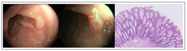
Figure 1 Colonic hyperplastic polyp with white light, narrow band imaging and hematoxylin-eosin stain. Histologic sample shows serrated architecture at the surface without glandular abnormalities. 

From the histological point of view, unlike normal epithelium of the colon, HP have a crypt convolved but not branched with a serrated profile of the epithelium at the level of the top half, thus giving the gland a starshaped in transverse section 14.

The subcategories (the most common "Microvesicular", which is characterized by an increased number of goblet cells, and the rarest "Mucin-poor") reported by WHO to date have not shown any clinical relevance but still show biomolecular differences 7.

Serrated sessile adenomas without dysplasia

Serrated sessile adenomas (SSA) without dysplasia appear in average larger than the HP in endoscopic imaging, since more than half have a size greater than 5 mm and approximately 15-20% are larger than 10 mm; these lesions are often covered with mucus, flat, with a yellowish/reddish or similar to the surrounding mucosa and found predominantly upstream of the splenic flexure (about 75%). These lesions constitute about 20% of all serrated lesions and about 10% of all colonic polyps 14,43.

From the histological view, unlike the HP, the architecture of the crypt has more jagged profile and the extension of this phenomenon is up to the basal half; SSA often appear with dilated crypts, extended laterally and behind the muscularis mucosae, forming inverted T or L-shaped crypts. This framework gives to the glands, a star-shaped and enlarged appearance compared to that of HP 44,45.

Serrated sessile adenomas with dysplasia (mixed polyps)

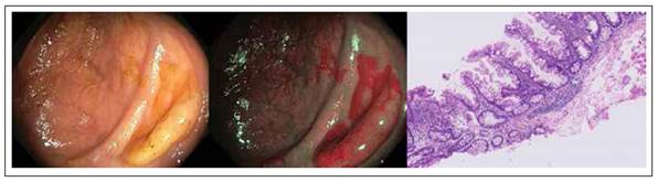
The endoscopic appearance of mixed polyps (MP) is similar to that of the SSA, as well as the pit pattern and distribution in the colon. Such lesions are about 4% of all serrated lesions and about 2% of all colonic polyps 43.

However, from the histological view, MP show areas reproducing the framework of the SSA and others with recognizable dysplasia, recalling the adenomatous structure 42 (Figure 2).

Figure 2 Colonic serrated sessile adenoma polyp with white light, narrow band imaging and hematoxylin-eosin stain. Histologic sample shows basal dilation of the crypts. 

Traditional serrated adenomas

Traditional serrated adenomas (TSA) endoscopically appear more similar to adenomas than to serrated polyps (Figure 3). In fact, they are most often reddish, sessile or pedunculated. These lesions constitute about 2% of all serrated lesions and 1% of all colonic polyps 34.

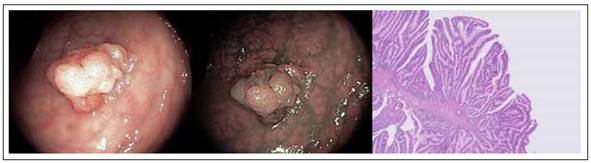
Figure 3 Colonic Traditional serrated adenomas (TSA) with white light, narrow band imaging and hematoxylin-eosin stain. Histologic sample shows protuberant villiform growth pattern with slit-like serrations and ectopic crypt formations. 

From the histological view, TSA have the same architecture of the crypt of the other serrated lesions but with cytologic aspect of widespread dysplasia (hyperchromasia of nuclei, elongation and stratification of nuclei, prominent nucleoli, hypereosinophilic cytoplasm). In cross section this framework gives to the gland a form more typically adenomatous with a pit pattern III according to Kudo classification 45,46.

WASP classification

Thanks to the novel technological improvements, in particular Narrow Band Imaging (NBI), the Workgroup on Serrated Polyps and Polyposis (WASP) has defined the so-called WASP classification 47, which can be used to perform a differential diagnosis between hyperplastic and adenomatous/serrated polyps. This classification uses as first step the NICE classification 47 to differentiate between type 1 polyps, which appear like same or lighter than background; none, or isolated lacy vessels may be present coursing across the lesion and dark or white spots of uniform size, or homogeneous absence of pattern) and type 2 polyps (browner relative to background, brown vessels surrounding white structures and oval, tubular or branched white structures surrounded by brown vessels). Then, the presence of 2 of the following ‘SSL-like features’ (i.e. clouded surface, indistinctive border, irregular shapes and dark spots inside crypts) strongly suggest the presence of SSA.

Risk Factors

The risk of serrated polyps, such for adenomatous polyps, is linked to environmental factors, related to lifestyle and diet. The factors causing an increase of the cancerogenic risk are excessive alcohol consumption, poor intake of folate and obesity 48,49. In the other hand, protective factors are regular use of anti-inflammatory drugs, hormone replacement therapy with estrogen and high calcium intake 48.

The effect of smoking cigarettes constitutes a real paradox, because it is significantly associated with an increased risk of serrated lesions but much less regarding to "serrated" colorectal neoplasms 48,50-53.

Age has been shown to be a factor that does not affect the risk of developing cancer even if the serrated colonic lesions appear earlier than adenomas 48,54,55. Sex, typically a risk factor for adenomatous polyps, increase the risk for SSA and for TSA; the male: female ratio to find a serrated neoplasia is of 1.9:1 56.

Endoscopic management

As all potential oncological lesions, the correct clinical management of serrated lesions is to recognize them during the colonoscopy and subsequently remove them, ideally in-bloc resection, then refer the sample to histological analysis. This approach is common to both adenomas and serrated lesions and is based on the concept that the removal of precancerous lesions means a significant reduction in mortality for colorectal cancer. This hypothesis is true only for the left colon: in fact, for the right colon the data in the literature up until now do not demonstrate a significant gain in terms of cancer mortality regarding the use of colonoscopy as a diagnostic and therapeutic method for such lesions 57.

Among the causes, it should be considered the presence especially in the right colon of SSA; such lesions, as previously illustrated, are difficult to diagnose due to the similar color with the surrounding mucosa and the capacity to be covered by mucus 14,43. In this regard, emerge more strongly as significant parameter of quality in colonoscopy, the rate of detection of serrated polyps which, together with that of adenomas, certify to the endoscopist of specific capacity in performing endoscopic exams with a lower miss rate of interval tumors 58. This parameter would stand at above 5% in the upstream portions of the splenic flexure 59-61, (the rate of detection of adenomas should be higher than 20% 62-64).

This assumption justifies the need of novel technology able to improve the detection of such lesions. Some early data investigated the role of Endocuff 65, which is a device composed of a soft, cylindrical, polymer with flexible circumferential projections that is mounted onto the distal tip of the endoscope. During the withdrawing, the hinged projections of this device flatten the colonic mucosal folds and increase the visualization of the colonic mucosa. A recent study by Baek et al. 66 showed that Endocuff assisted colonoscopy was able to detect more SSA than standard colonoscopy (59 vs 8; p ≤ 0.0001) and significantly increased the SSA detection rate (15% vs 3%; p ≤ 0.0001).

Another potentially useful technology is the G-EYE 67 which is a novel device comprises a reusable balloon integrated on a conventional colonoscope which during the withdrawal phase is partially inflated. This can potentially flatten the colonic folds and stabilize the colonoscope avoiding bowel slippage. In a large randomized controlled study, Shirin et al.67 showed that G-EYE was able to significantly improve the detection of SSA (3 vs 20; p = 0.0026). Finally, a recent meta-analysis of Atkinson et al. (68), including 11 studies, evaluated the role of NBI in the detection of SSA. Results showed that NBI was able to detected more non-adenomatous polyps (OR, 1.24; 95% CI, 1.06-1.44; p = 0.008) and flat polyps (OR, 1.24; 95% CI, 1.02-1.51; p = 0.03) than white light endoscopy.

Serrated polyposis syndrome (SPS)

Definition and diagnostic criteria

Serrated polyposis syndrome (Syndrome-SPS), a condition originally known as hyperplastic polyposis, is characterized by a high number of colorectal polyps with serrated architecture. As the SPS is poor known and poor understood condition, it presents a high risk for personal and family CRC.

The diagnostic criteria of the SPS were recently redefined by ESGE guidelines 69: (1) at least 5 polyps resected proximal to the sigmoid colon, 2 of which> 10 mm, or (2) > 20 serrated lesions of any size distributed in the entire colon.

Clinical Features

The clinical characteristics of patients with SPS have been mostly defined on the basis of case-series 70-72. SPS has no predominance of sex and the average age of diagnosis is about 55 years. SPS has long been considered genetic disease but the pattern of inheritance remains unknown: mechanisms suggested both recessive and dominant autosomal inheritance.

Moreover, 10-50% of patients with SPS have a family history of CRC and there is an increased risk of CRC in their first-degree relatives compared to general population 71-74. It’s important to point out that the conventional adenomas may coexist with serrated polyps in patients with SPS 70-72.

Environmental factors (obesity, smoking, increased dietary intake of red meat and fat) may be partially responsible for the onset of serrated lesions (not necessarily SPS) in the left colon. In the right colon, folate intake and family history of polyps and aspirin would have a protective role 75.

Risk of cancer and recommendations for treatment and surveillance

SPS has a high risk of CRC in subjects between 50 and 60 years 70. In published series, approximately 25%70% of patients with SPS had the CRC at the time of diagnosis or the next follow-up 74,76,77. There is a high incidence of synchronous cancer 76 and the CRC shows greater tendency to be localized to the proximal colon in patient with SPS 71.

First-degree relatives of patients with SPS have a higher risk of CRC than the general population 73.

Regular screening colonoscopy should be performed in order to remove potential premalignant lesions and bearing in mind that the detection of the serrated polyp is often difficult (often sessile or flat lesions). The fecal occult blood plays a minor role because serrated lesions have less bleeding tendency than the adenomas.

Recommendations on surveillance strategies

Up to date, several studies showed how traditional serrated adenoma, serrated polyp ≥ 10mm and serrated polyp with dysplasia are similar to conventional adenomas in terms of metachronous advanced neoplasia or CRC risks 78-80. In detail, one population-based randomized study on 12 955 patients screened with flexible sigmoidoscopy 81 and a recent retrospective study 82 evaluating 122 899 patients with 10 years of follow-up showed higher Hazard Rate of metachronous CRC of 4.2 (95%CI 1.3 - 13.3) and 3.35 (95 %CI 1.37 - 8.15) Therefore, these lesions (i.e. traditional serrated adenoma, serrated polyp ≥ 10mm and serrated polyp with dysplasia) need endoscopic surveillance with colonoscopy at 3 years according to the recent ESGE guidelines on post-polypectomy surveillance 83.

Also SPS has been widely discussed in recent national and international guidelines (i.e. USA 59, UK 84, Europe 69, Japan 85, Korean 86 and Australian 87). As above mentioned, SPS patients showed a higher risk of CRC. Therefore, ESGE recommends 69 endoscopic removal of all polyps ≥ 5mm and all polyps of any size with optical suspicion of dysplasia in individuals with serrated polyposis syndrome before and after entering surveillance. Regarding the surveillance, ESGE recommends 69 interval of post-polypectomy surveillance at 1 year after ≥1 advanced polyp or ≥ 5 nonadvanced clinically relevant polyps and at 2 years after no advanced polyps or < 5 nonadvanced clinically relevant polyps.

Since several studies showed an increased incidence of CRC in relatives of patients with SPS 88-90. Therefore, follow-up of these individuals is suggested by ESGE guidelines with colonoscopy from the age of 45 years. In case positive findings, the patient will follow the abovementioned post-polypectomy surveillance. Otherwise, colonoscopy should be performed every 5 years 83.

CONCLUSION

This literature review showed that the serrated lesions are a cauldron with very heterogeneous genetic and biological characteristics causing important clinical consequences.

Such lesions are in fact now recognized as precancerous lesions. This statement is true both for the sporadic forms that familiar ones, and for this reason the scientific world showing more and more interest in knowing the biological processes that underlie them to neoplastic transformation, especially for their significant impact in carcinogenesis in the right colon. Data of the literature show as it’s increasing necessity to have further internationally accepted guidelines regarding the clinical and endoscopic management of these lesions.

REFERENCES

1. Jemal A, Siegel R, Ward E, Murray T, Xu J, Smigal C, Thun MJ. Cancer statistics, 2006. CA Cancer J Clin. 2006;56(2):106- 30. doi: 10.3322/canjclin.56.2.106. [ Links ]

2. Cho KR, Vogelstein B. Genetic alterations in the adenoma- -carcinoma sequence. Cancer. 1992;70(6 Suppl):1727-31. [ Links ]

3. Schmieden W, Westhues H. Zur Klinik und Pathologie der Dickdarmpolypell und deren klinischen und pathologischanatomischen Beziehungen zum Dickdarmkarzinom. Deutsche Ztschr f Chir. 1927;202:1-124. [ Links ]

4. Goldman H, Ming S, Hickock DF. Nature and significance of hyperplastic polyps of the human colon. Arch Pathol. 1970;89(4):349-54. [ Links ]

5. Urbanski SJ, Kossakowska AE, Marcon N, Bruce WR. Mixed hyperplastic adenomatous polyps--an underdiagnosed entity. Report of a case of adenocarcinoma arising within a mixed hyperplastic adenomatous polyp. Am J Surg Pathol. 1984;8(7):551-6. [ Links ]

6. Longacre TA, Fenoglio-Preiser CM. Mixed hyperplastic adenomatous polyps/serrated adenomas. A distinct form of colorectal neoplasia. Am J Surg Pathol. 1990;14(6):524-37. doi: 10.1097/00000478-199006000-00003. [ Links ]

7. Torlakovic E, Skovlund E, Snover DC, Torlakovic G, Nesland JM. Morphologic reappraisal of serrated colorectal polyps. Am J Surg Pathol. 2003;27(1):65-81. doi: 10.1097/00000478-200301000-00008. [ Links ]

8. Snover DC, Ahnen DJ, Burt RW, Odze RD. Serrated polyps of the colon and rectum and serrated polyposis. In: Bosman FT, Carneiro F, Hruban RH, Theise ND, editors. WHO classification of tumours of the digestive system. Lyon, France: IARC; 2010. p. 160-5. [ Links ]

9. Nagtegaal ID, Odze RD, Klimstra D, Paradis V, Rugge M, Schirmacher P, et al. The 2019 WHO classification of tumours of the digestive system. Histopathology. 2020;76(2):182- 188. doi: 10.1111/his.13975. [ Links ]

10. Crockett SD, Nagtegaal ID. Terminology, Molecular Features, Epidemiology, and Management of Serrated Colorectal Neoplasia. Gastroenterology. 2019;157(4):949-966.e4. doi: 10.1053/j.gastro.2019.06.041. [ Links ]

11. Szylberg L, Janiczek M, Popiel A, Marszalek A. Serrated polyps and their alternative pathway to the colorectal cancer: a systematic review. Gastroenterol Res Pract. 2015;2015:573814. doi: 10.1155/2015/573814. [ Links ]

12. Rajagopalan H, Bardelli A, Lengauer C, Kinzler KW, Vogelstein B, Velculescu VE. Tumorigenesis: RAF/RAS oncogenes and mismatch-repair status. Nature. 2002;418(6901):934. doi: 10.1038/418934a. [ Links ]

13. Yang S, Farraye FA, Mack C, Posnik O, O&apos;Brien MJ. BRAF and KRAS Mutations in hyperplastic polyps and serrated adenomas of the colorectum: relationship to histology and CpG island methylation status. Am J Surg Pathol. 2004;28(11):1452-9. doi: 10.1097/01.pas.0000141404.56839.6a. [ Links ]

14. O&apos;Brien MJ. Hyperplastic and serrated polyps of the colorectum. Gastroenterol Clin North Am. 2007;36(4):947- 68, viii. doi: 10.1016/j.gtc.2007.08.007. [ Links ]

15. Jass JR, Baker K, Zlobec I, Higuchi T, Barker M, Buchanan D, et al. Advanced colorectal polyps with the molecular and morphological features of serrated polyps and adenomas: concept of a &apos;fusion&apos; pathway to colorectal cancer. Histopathology. 2006;49(2):121-31. doi: 10.1111/j.1365-2559.2006.02466.x. [ Links ]

16. Konishi K, Yamochi T, Makino R, Kaneko K, Yamamoto T, Nozawa H, et al. Molecular differences between sporadic serrated and conventional colorectal adenomas. Clin Cancer Res. 2004;10(9):3082-90. doi: 10.1158/1078-0432.ccr-03-0334. [ Links ]

17. Dong SM, Lee EJ, Jeon ES, Park CK, Kim KM. Progressive methylation during the serrated neoplasia pathway of the colorectum. Mod Pathol. 2005;18(2):170-8. doi: 10.1038/modpathol.3800261. [ Links ]

18. McGivern A, Wynter CV, Whitehall VL, Kambara T, Spring KJ, Walsh MD, et al. Promoter hypermethylation frequency and BRAF mutations distinguish hereditary non-polyposis colon cancer from sporadic MSI-H colon cancer. Fam Cancer. 2004;3(2):101-7. doi: 10.1023/B:FAME.0000039861.30651.c8. [ Links ]

19. Erhardt P, Schremser EJ, Cooper GM. B-Raf inhibits programmed cell death downstream of cytochrome c release from mitochondria by activating the MEK/Erk pathway. Mol Cell Biol. 1999;19(8):5308-15. doi: 10.1128/MCB.19.8.5308. [ Links ]

20. Hörkkö TT, Mäkinen MJ. Colorectal proliferation and apoptosis in serrated versus conventional adenomacarcinoma pathway: growth, progression and survival. Scand J Gastroenterol. 2003;38(12):1241-1248. doi: 10.1080/00365520310006603. [ Links ]

21. Jubb AM, Bell SM, Quirke P. Methylation and colorectal cancer. J Pathol. 2001;195(1):111-134. doi: 10.1002/path.923. [ Links ]

22. Kambara T, Simms LA, Whitehall VL, Spring KJ, Wynter CV, Walsh MD, et al. BRAF mutation is associated with DNA methylation in serrated polyps and cancers of the colorectum. Gut. 2004;53(8):1137-44. doi: 10.1136/gut.2003.037671. [ Links ]

23. Ogino S, Kawasaki T, Kirkner GJ, Ogawa A, Dorfman I, Loda M, et al. Down-regulation of p21 (CDKN1A/CIP1) is inversely associated with microsatellite instability and CpG island methylator phenotype (CIMP) in colorectal cancer. J Pathol. 2006;210(2):147-54. doi: 10.1002/path.2030. [ Links ]

24. Park SJ, Rashid A, Lee JH, Kim SG, Hamilton SR, Wu TT. Frequent CpG island methylation in serrated adenomas of the colorectum. Am J Pathol. 2003;162(3):815-22. doi: 10.1016/S0002-9440(10)63878-3. [ Links ]

25. Chan AO, Issa JP, Morris JS, Hamilton SR, Rashid A. Concordant CpG island methylation in hyperplastic polyposis. Am J Pathol. 2002;160(2):529-36. doi: 10.1016/S0002-9440(10)64872-9. [ Links ]

26. Kondo Y, Issa JP. Epigenetic changes in colorectal cancer. Cancer Metastasis Rev. 2004;23(1-2):29-39. doi: 10.1023/a:1025806911782. [ Links ]

27. van Rijnsoever M, Grieu F, Elsaleh H, Joseph D, Iacopetta B. Characterisation of colorectal cancers showing hypermethylation at multiple CpG islands. Gut. 2002;51(6):797-802. doi: 10.1136/gut.51.6.797. [ Links ]

28. Jass JR. Serrated adenoma of the colorectum and the DNAmethylator phenotype. Nat Clin Pract Oncol. 2005;2(8):398- 405. doi: 10.1038/ncponc0248. [ Links ]

29. Minoo P, Baker K, Goswami R, Chong G, Foulkes WD, Ruszkiewicz AR, et al. Extensive DNA methylation in normal colorectal mucosa in hyperplastic polyposis. Gut. 2006;55(10):1467-74. doi: 10.1136/gut.2005.082859. [ Links ]

30. Kawakami K, Ruszkiewicz A, Bennett G, Moore J, Grieu F, Watanabe G, et al. DNA hypermethylation in the normal colonic mucosa of patients with colorectal cancer. Br J Cancer. 2006;94(4):593-8. doi: 10.1038/sj.bjc.6602940. [ Links ]

31. Young J, Simms LA, Biden KG, Wynter C, Whitehall V, Karamatic R, et al. Features of colorectal cancers with high-level microsatellite instability occurring in familial and sporadic settings: parallel pathways of tumorigenesis. Am J Pathol. 2001;159(6):2107-16. doi: 10.1016/S0002-9440(10)63062-3. [ Links ]

32. Deng G, Bell I, Crawley S, Gum J, Terdiman JP, Allen BA, et al. BRAF mutation is frequently present in sporadic colorectal cancer with methylated hMLH1, but not in hereditary nonpolyposis colorectal cancer. Clin Cancer Res. 2004;10(1 Pt 1):191-5. doi: 10.1158/1078-0432.ccr-1118-3.5. [ Links ]

33. Jass JR. HNPCC and sporadic MSI-H colorectal cancer: a review of the morphological similarities and differences. Fam Cancer. 2004;3(2):93-100. doi: 10.1023/B:FAME.0000039849.86008.b7. [ Links ]

34. Huang CS, O&apos;brien MJ, Yang S, Farraye FA. Hyperplastic polyps, serrated adenomas, and the serrated polyp neoplasia pathway. Am J Gastroenterol. 2004;99(11):2242-55. doi: 10.1111/j.1572-0241.2004.40131.x. [ Links ]

35. O&apos;Brien MJ, Yang S, Mack C, Xu H, Huang CS, Mulcahy E, et al. Comparison of microsatellite instability, CpG island methylation phenotype, BRAF and KRAS status in serrated polyps and traditional adenomas indicates separate pathways to distinct colorectal carcinoma end points. Am J Surg Pathol. 2006;30(12):1491-501. doi: 10.1097/01.pas.0000213313.36306.85. [ Links ]

36. Snover DC, Batts KP. Serrated Colorectal Neoplasia. Surg Pathol Clin. 2010;3(2):207-40. doi: 10.1016/j.path.2010.05.001. [ Links ]

37. Waye JD, Bilotta JJ. Rectal hyperplastic polyps: now you see them, now you don&apos;t--a differential point. Am J Gastroenterol. 1990;85(12):1557-9. [ Links ]

38. Chapuis PH, Dent OF, Goulston KJ. Clinical accuracy in the diagnosis of small polyps using the flexible fiberoptic sigmoidoscope. Dis Colon Rectum. 1982;25(7):669-72. doi: 10.1007/BF02629538. [ Links ]

39. Norfleet RG, Ryan ME, Wyman JB. Adenomatous and hyperplastic polyps cannot be reliably distinguished by their appearance through the fiberoptic sigmoidoscope. Dig Dis Sci. 1988;33(9):1175-7. doi: 10.1007/BF01535796. [ Links ]

40. Imperiale TF, Wagner DR, Lin CY, Larkin GN, Rogge JD, Ransohoff DF. Risk of advanced proximal neoplasms in asymptomatic adults according to the distal colorectal findings. N Engl J Med. 2000;343(3):169-74. doi: 10.1056/NEJM200007203430302. [ Links ]

41. Imperiale TF, Wagner DR, Lin CY, Larkin GN, Rogge JD, Ransohoff DF. Results of screening colonoscopy among persons 40 to 49 years of age. N Engl J Med. 2002;346(23):1781-5. doi: 10.1056/NEJM200206063462304. [ Links ]

42. Mäkinen MJ. Colorectal serrated adenocarcinoma. Histopathology. 2007;50(1):131-50. doi: 10.1111/j.1365-2559.2006.02548.x. [ Links ]

43. Spring KJ, Zhao ZZ, Karamatic R, Walsh MD, Whitehall VL, Pike T, et al. High prevalence of sessile serrated adenomas with BRAF mutations: a prospective study of patients undergoing colonoscopy. Gastroenterology. 2006;131(5):1400-7. doi: 10.1053/j.gastro.2006.08.038. [ Links ]

44. Peyssonnaux C, Eychène A. The Raf/MEK/ERK pathway: new concepts of activation. Biol Cell. 2001;93(1-2):53-62. doi: 10.1016/s0248-4900(01)01125-x. [ Links ]

45. Mansoor I. Clinicopathological analysis of serrated adenomas of the colorectum. The Internet Journal of Pathology. 2003;2(2). [ Links ]

46. Li SC, Burgart L. Histopathology of serrated adenoma, its variants, and differentiation from conventional adenomatous and hyperplastic polyps. Arch Pathol Lab Med. 2007;131(3):440-5. doi: 10.5858/2007-131-440-HOSAIV. [ Links ]

47. Hewett DG, Kaltenbach T, Sano Y, Tanaka S, Saunders BP, Ponchon T, et al. Validation of a simple classification system for endoscopic diagnosis of small colorectal polyps using narrow-band imaging. Gastroenterology. 2012;143(3):599- 607.e1. doi: 10.1053/j.gastro.2012.05.006. [ Links ]

48. Morimoto LM, Newcomb PA, Ulrich CM, Bostick RM, Lais CJ, Potter JD. Risk factors for hyperplastic and adenomatous polyps: evidence for malignant potential? Cancer Epidemiol Biomarkers Prev. 2002;11(10 Pt 1):1012-8. [ Links ]

49. Kearney J, Giovannucci E, Rimm EB, Stampfer MJ, Colditz GA, Ascherio A, et al. Diet, alcohol, and smoking and the occurrence of hyperplastic polyps of the colon and rectum (United States). Cancer Causes Control. 1995;6(1):45-56. doi: 10.1007/BF00051680. [ Links ]

50. Young J. Serrated neoplasia of the colorectum and cigarette smoking. Gastroenterology. 2008;135(1):323-324; author reply 324. doi: 10.1053/j.gastro.2008.03.080. [ Links ]

51. Ji BT, Weissfeld JL, Chow WH, Huang WY, Schoen RE, Hayes RB. Tobacco smoking and colorectal hyperplastic and adenomatous polyps. Cancer Epidemiol Biomarkers Prev. 2006;15(5):897-901. doi: 10.1158/1055-9965.EPI-05-0883. [ Links ]

52. Shrubsole MJ, Wu H, Ness RM, Shyr Y, Smalley WE, Zheng W. Alcohol drinking, cigarette smoking, and risk of colorectal adenomatous and hyperplastic polyps. Am J Epidemiol. 2008;167(9):1050-8. doi: 10.1093/aje/kwm400. [ Links ]

53. Anderson JC, Pleau DC, Rajan TV, Protiva P, Swede H, Brenner B, et al. Increased frequency of serrated aberrant crypt foci among smokers. Am J Gastroenterol. 2010;105(7):1648- 54. doi: 10.1038/ajg.2010.109. [ Links ]

54. Clark JC, Collan Y, Eide TJ, Estève J, Ewen S, Gibbs NM, et al. Prevalence of polyps in an autopsy series from areas with varying incidence of large-bowel cancer. Int J Cancer. 1985;36(2):179-86. doi: 10.1002/ijc.2910360209. [ Links ]

55. Isbister WH. Hyperplastic polyps. ANZ J Surg. 1993;63(3):175-180. [ Links ]

56. Tuppurainen K, Makinen JM, Junttila O, Liakka A, Kyllonen AP, Tuominen H, et al. Morphology and microsatellite instability in sporadic serrated and non-serrated colorectal cancer. J Pathol. 2005;207(3):285-94. doi: 10.1002/path.1850. [ Links ]

57. Rex DK, Bond JH, Winawer S, Levin TR, Burt RW, Johnson DA, et al. Quality in the technical performance of colonoscopy and the continuous quality improvement process for colonoscopy: recommendations of the U.S. Multi-Society Task Force on Colorectal Cancer. Am J Gastroenterol. 2002;97(6):1296- 308. doi: 10.1111/j.1572-0241.2002.05812.x. [ Links ]

58. Pabby A, Schoen RE, Weissfeld JL, Burt R, Kikendall JW, Lance P, et al. Analysis of colorectal cancer occurrence during surveillance colonoscopy in the dietary Polyp Prevention Trial. Gastrointest Endosc. 2005;61(3):385-91. doi: 10.1016/s0016-5107(04)02765-8. [ Links ]

59. Lieberman DA, Rex DK, Winawer SJ, Giardiello FM, Johnson DA, Levin TR. Guidelines for colonoscopy surveillance after screening and polypectomy: a consensus update by the US Multi-Society Task Force on Colorectal Cancer. Gastroenterology. 2012;143(3):844-857. doi: 10.1053/j.gastro.2012.06.001. [ Links ]

60. Schreiner MA, Weiss DG, Lieberman DA. Proximal and large hyperplastic and nondysplastic serrated polyps detected by colonoscopy are associated with neoplasia. Gastroenterology. 2010;139(5):1497-502. doi: 10.1053/j.gastro.2010.06.074. [ Links ]

61. Liang J, Kalady MF, Appau K, Church J. Serrated polyp detection rate during screening colonoscopy. Colorectal Dis. 2012;14(11):1323-7. doi: 10.1111/j.1463- 1318.2012.03017.x. [ Links ]

62. Barclay RL, Vicari JJ, Doughty AS, Johanson JF, Greenlaw RL. Colonoscopic withdrawal times and adenoma detection during screening colonoscopy. N Engl J Med. 2006;355(24):2533-41. doi: 10.1056/NEJMoa055498. [ Links ]

63. Millan MS, Gross P, Manilich E, Church JM. Adenoma detection rate: the real indicator of quality in colonoscopy. Dis Colon Rectum. 2008;51(8):1217-20. doi: 10.1007/s10350-008-9315-3. [ Links ]

64. Church J. Adenoma detection rate and the quality of colonoscopy: the sword has two edges. Dis Colon Rectum. 2008;51(5):520-3. doi: 10.1007/s10350-008-9239-y. [ Links ]

65. Lenze F, Beyna T, Lenz P, Heinzow HS, Hengst K, Ullerich H. Endocuff-assisted colonoscopy: a new accessory to improve adenoma detection rate? Technical aspects and first clinical experiences. Endoscopy. 2014;46(7):610-4. doi: 10.1055/s-0034-1365446. [ Links ]

66. Baek MD, Jackson CS, Lunn J, Nguyen C, Shah NK, Serrao S, et al. Endocuff assisted colonoscopy significantly increases sessile serrated adenoma detection in veterans. J Gastrointest Oncol. 2017;8(4):636-642. doi: 10.21037/jgo.2017.03.07. [ Links ]

67. Shirin H, Shpak B, Epshtein J, Karstensen JG, Hoffman A, de Ridder R, Testoni PA, Ishaq S, Reddy DN, Gross SA, Neumann H, Goetz M, Abramowich D, Moshkowitz M, Mizrahi M, Vilmann P, Rey JW, Sanduleanu-Dascalescu S, Viale E, Chaudhari H, Pochapin MB, Yair M, Shnell M, Yaari S, Hendel JW, Teubner D, Bogie RMM, Notaristefano C, Simantov R, Gluck N, Israeli E, Stigaard T, Matalon S, Vilkin A, Benson A, Sloth S, Maliar A, Waizbard A, Jacob H, Thielsen P, Shachar E, Rochberger S, Hershcovici T, Plougmann JI, Braverman M, Tsvang E, Abedi AA, Brachman Y, Siersema PD, Kiesslich R. G-EYE colonoscopy is superior to standard colonoscopy for increasing adenoma detection rate: an international randomized controlled trial (with videos). Gastrointest Endosc. 2019 Mar;89(3):545-553. [ Links ]

68. Atkinson NSS, Ket S, Bassett P, Aponte D, De Aguiar S, Gupta N, et al. Narrow-Band Imaging for Detection of Neoplasia at Colonoscopy: A Meta-analysis of Data From Individual Patients in Randomized Controlled Trials. Gastroenterology. 2019;157(2):462-471. doi: 10.1053/j.gastro.2019.04.014. [ Links ]

69. van Leerdam ME, Roos VH, van Hooft JE, Dekker E, Jover R, Kaminski MF, et al. Endoscopic management of polyposis syndromes: European Society of Gastrointestinal Endoscopy (ESGE) Guideline. Endoscopy. 2019;51(9):877-895. doi: 10.1055/a-0965-0605. [ Links ]

70. Chow E, Lipton L, Lynch E, D&apos;Souza R, Aragona C, Hodgkin L, et al. Hyperplastic polyposis syndrome: phenotypic presentations and the role of MBD4 and MYH. Gastroenterology. 2006;131(1):30-9. doi: 10.1053/j.gastro.2006.03.046. [ Links ]

71. Rubio CA, Stemme S, Jaramillo E, Lindblom A. Hyperplastic polyposis coli syndrome and colorectal carcinoma. Endoscopy. 2006;38(3):266-70. doi: 10.1055/s-2006-925026. [ Links ]

72. Kalady MF, Jarrar A, Leach B, LaGuardia L, O&apos;Malley M, Eng C, et al. Defining phenotypes and cancer risk in hyperplastic polyposis syndrome. Dis Colon Rectum. 2011;54(2):164-70. doi: 10.1007/DCR.0b013e3181fd4c15. [ Links ]

73. Boparai KS, Reitsma JB, Lemmens V, van Os TA, Mathus- Vliegen EM, Koornstra JJ, et al. Increased colorectal cancer risk in first-degree relatives of patients with hyperplastic polyposis syndrome. Gut. 2010;59(9):1222-5. doi: 10.1136/gut.2009.200741. [ Links ]

74. Wallace K, Grau MV, Ahnen D, Snover DC, Robertson DJ, Mahnke D, Gui J, Barry EL, Summers RW, McKeown-Eyssen G, Haile RW, Baron JA. The association of lifestyle and dietary factors with the risk for serrated polyps of the colorectum. Cancer Epidemiol Biomarkers Prev. 2009;18(8):2310- 7. doi: 10.1158/1055-9965.EPI-09-0211. [ Links ]

75. Yeoman A, Young J, Arnold J, Jass J, Parry S. Hyperplastic polyposis in the New Zealand population: a condition associated with increased colorectal cancer risk and European ancestry. N Z Med J. 2007;120(1266):U2827. [ Links ]

76. Leggett B, Whitehall V. Role of the serrated pathway in colorectal cancer pathogenesis. Gastroenterology. 2010;138(6):2088-100. doi: 10.1053/j.gastro.2009.12.066. [ Links ]

77. Brooker JC, Saunders BP, Shah SG, Thapar CJ, Suzuki N, Williams CB. Treatment with argon plasma coagulation reduces recurrence after piecemeal resection of large sessile colonic polyps: a randomized trial and recommendations. Gastrointest Endosc. 2002;55(3):371-5. doi: 10.1067/mge.2002.121597. [ Links ]

78. Anderson JC, Butterly LF, Robinson CM, Weiss JE, Amos C, Srivastava A. Risk of Metachronous High-Risk Adenomas and Large Serrated Polyps in Individuals With Serrated Polyps on Index Colonoscopy: Data From the New Hampshire Colonoscopy Registry. Gastroenterology. 2018;154(1):117-127. e2. doi: 10.1053/j.gastro.2017.09.011. [ Links ]

79. Erichsen R, Baron JA, Hamilton-Dutoit SJ, Snover DC, Torlakovic EE, Pedersen L, et al. Increased Risk of Colorectal Cancer Development Among Patients With Serrated Polyps. Gastroenterology. 2016;150(4):895-902.e5. doi: 10.1053/j.gastro.2015.11.046. [ Links ]

80. Yoon JY, Kim HT, Hong SP, Kim HG, Kim JO, Yang DH, et al. High-risk metachronous polyps are more frequent in patients with traditional serrated adenomas than in patients with conventional adenomas: a multicenter prospective study. Gastrointest Endosc. 2015;82(6):1087-93.e3. doi: 10.1016/j.gie.2015.05.016. [ Links ]

81. Holme Ø, Bretthauer M, Eide TJ, Løberg EM, Grzyb K, Løberg M, et al. Long-term risk of colorectal cancer in individuals with serrated polyps. Gut. 2015;64(6):929-36. doi: 10.1136/gutjnl-2014-307793. [ Links ]

82. He X, Hang D, Wu K, Nayor J, Drew DA, Giovannucci EL, et al. Long-term Risk of Colorectal Cancer After Removal of Conventional Adenomas and Serrated Polyps. Gastroenterology. 2020;158(4):852-861.e4. doi: 10.1053/j.gastro.2019.06.039. [ Links ]

83. Hassan C, Antonelli G, Dumonceau JM, Regula J, Bretthauer M, Chaussade S, et al. Post-polypectomy colonoscopy surveillance: European Society of Gastrointestinal Endoscopy (ESGE) Guideline - Update 2020. Endoscopy. 2020;52(8):687-700. doi: 10.1055/a-1185-3109. [ Links ]

84. East JE, Atkin WS, Bateman AC, Clark SK, Dolwani S, Ket SN, et al. British Society of Gastroenterology position statement on serrated polyps in the colon and rectum. Gut. 2017;66(7):1181-1196. doi: 10.1136/gutjnl-2017-314005. [ Links ]

85. Tanaka S, Saitoh Y, Matsuda T, Igarashi M, Matsumoto T, Iwao Y, et al. Evidence-based clinical practice guidelines for management of colorectal polyps. J Gastroenterol. 2015;50(3):252-60. doi: 10.1007/s00535-014-1021-4. [ Links ]

86. Yang DH, Hong SN, Kim YH, Hong SP, Shin SJ, Kim SE, et al, Surveillance and Management. Korean guidelines for postpolypectomy colonoscopy surveillance. Clin Endosc. 2012;45(1):44-61. doi: 10.5946/ce.2012.45.1.44. [ Links ]

87. Cancer Council Australia Colonoscopy Surveillance Working Party. Clinical Practice Guidelines for Surveillance Colonoscopy-in adenoma follow-up; following curative resection of colorectal cancer; and for cancer surveillance in inflammatory bowel disease. Sydney: Cancer Council Australia; 2011. [ Links ]

88. Oquiñena S, Guerra A, Pueyo A, Eguaras J, Montes M, Razquin S, et al. Serrated polyposis: prospective study of firstdegree relatives. Eur J Gastroenterol Hepatol. 2013;25(1):28- 32. doi: 10.1097/MEG.0b013e3283598506. [ Links ]

89. Caetano AC, Ferreira H, Soares J, Ferreira A, Gonçalves R, Rolanda C. Phenotypic characterization and familial risk in hyperplastic polyposis syndrome. Scand J Gastroenterol. 2013;48(10):1166-72. doi: 10.3109/00365521.2013.830329. [ Links ]

90. Lage P, Cravo M, Sousa R, Chaves P, Salazar M, Fonseca R, et al. Management of Portuguese patients with hyperplastic polyposis and screening of at-risk first-degree relatives: a contribution for future guidelines based on a clinical study. Am J Gastroenterol. 2004;99(9):1779-84. doi: 10.1111/j.1572-0241.2004.30178.x. [ Links ]

Disclosure statements: The authors have no conflicts of interest to declare.

Funding sources: The authors have no funding sources related to this article.

Received: May 26, 2021; Accepted: October 25, 2021

Correspondence: Rodrigo Mansilla-Vivar Gastrointestinal endoscopy Unit, Hospital Puerto Montt, Chile. Gastrointestinal Endoscopy Research Group, IIS Hospital La Fe, Valencia, Spain. Los Aromos 65, ZC 5480000 Phone: +56989061010 Puerto Montt, Chile E-mail: rmansillavivar@gmail.com

Author contributions: Rodrigo Mansilla-Vivar, Carlo Petruzzellis, Nicola Olivari, Sebastian Manuel Milluzzo and Alessandra Grassano wrote the manuscript, Pietro Cesari and Cristiano Spada revised the manuscript for important intellectual content, Fausto Zorzi revised all the data related with histology. All authors have read and approved the final version to be published.

Creative Commons License This is an open-access article distributed under the terms of the Creative Commons Attribution License