


 Espanhol (pdf)
 Artigo em XML
 Referências do artigo
 Enviar este artigo por email
 Citado por SciELO
  Similares em
SciELO
Similares em
SciELO  uBio
uBio 
 Permalink
INTRODUCCIÓN
Squamous cell carcinoma (SCC) is an invasive skin neoplasm that can originate in several regions, with a predisposition for areas with less skin pigmentation or without hair (Taylor and Haldorson, 2012; Rabbers et al., 2014). Secondary bacterial contamination can happen because it is a cutaneous tumour and therefore it is possible to observe the presence of purulent content on its surface (Ramos et al., 2007).
Initially, this neoplasm can appear as a solar dermatosis, and with its evolution it forms erythema, peeling and oedema, consequently, crusts are formed, altering the epidermis, ulcerating, and resulting in a cauliflower aspect. The diagnosis is obtained through anamnesis, animal history, aspect of the lesion; however, diagnostic confirmation can only be performed through cytological or histopathological exams (van den Top et al., 2008, 2010).
The differential diagnosis must be made with sarcoid, pythiosis, habronemosis, exuberant granulation tissue, strephano- flariosis, papilloma and fibropapiloma through laboratory tests (McGavin and Zachary, 2009; Coelho et al., 2012). When diagnosed early, the indicated therapy for horses is surgery (Fernandes, 2007). Thus, the early diagnosis of this disease and the institution of adequate treatment are essential for therapeutic success, especially in places where there are no adequate veterinary medical services. Therefore, the aim of this study was to report a case of squamous cell carcinoma that occurred in a horse diagnosed through histopathological examination.
CASE REPORT
Thew owner of a crossbred female, brown horse, approximately 12 years old, resident in the riverside community of Ipanema, on the banks of the Amazon River called the veterinary hospital of the Centro Universitário da Amazônia (UNAMA) requesting veterinary assistance. The owner could not take the animal to UNAMA due to the difficulties of transport in rural areas and the low zootechnical value of the animal.
According to the tutor’s report, the ani- mal had a lesion in the vulva region that had evolved for six months and had not responded to previous treatment with an association of therapy with antibiotics and corticoids. This lesion was treated as a myiasis, as there were many larvae in the lesion, however, after the treatment of this disease, no clinical regression was observed.
During the semiological examination, apathy, dehydration and severe cachexia were observed. On physical examination, a slight inflammation of the vulvar lips, in the region below the tail and in the perineum was observed, with erythema, oedema and desquamation. Besides, productive, granulomatous neoformation, with increased volume and with cauliflower appearance that bled easily was observed. The tumours have a moist appearance and ulcerations, a foul smell and bloody secretion, in an area with a break in the skin (Figure 1).
Three irregular fragments measuring an average of 2.0 cm in diameter were collected and placed in a 50 ml falcon tube with 10% formaldehyde. The collected material was sent to a private outsourced laboratory, where it was processed for histopathological examination. The samples were embedded in paraffin, cut to 5 µm slides and stained with haematoxylin-eosin (HE).
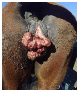
Figure 1 Image of the tumour, with increased volume in the vulva region, moist and ulcerated appearance, in addition to bloody secretion at the site
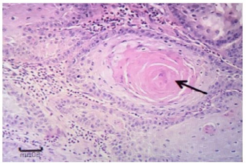
The outsourced laboratory did not provide a photomicrograph of the histopathology; however, the microscopic description revealed neoplastic proliferation of epithelial cells, supported by moderate fibrovascular stroma, forming islands and trabeculae. The cells were oval to polyhedral, juxtaposed, interconnected by desmosomal junctions, with ample and eosinophilic cytoplasm. Nuclei were oval, with loose chromatin and 1 to 3 prominent nucleoli. In some areas, abrupt keratinization of neoplastic cells was observed, with dyskeratosis and corneal pearl formation (Figure 2).
There was moderate anisokaryosis and anisocytosis and two mitotic figures in 10 higher power fields. Two microscopic readings were performed and there was an agreement of various veterinary pathologists regarding the results of the histopathological findings, which concluded that they were compatible with squamous cell carcinoma.
DISCUSSION
The mare had a lesion in the vulvar region compatible with SCC. Taylor and Haldorson (2012) indicated that about 13% of the cases of SCC in horses involves the external genitalia. In addition, it is noteworthy that when occurs in the vulvar region have a proliferative form, which can reach surrounding tissues reaching the vaginal vestibule.
The characteristics of the lesions in the vulva region were in accordance with other authors (Brinsko, 1998; Gross et al., 2005; van den Top et al., 2011), who describe the lesions as small ulcerations or superficial plaques of difficult healing. Valentine (2006) indicates that that the location with the greatest predisposition for the appearance of SCC in horses are the region of the penis and / or foreskin, ocular region, vulva and perianal region and skin.
The histopathological exam is required to reach a conclusive diagnosis (McGavin and Zachary, 2009; Zachary et al., 2012). The histological analysis revealed neoplastic proliferation of epithelial cells, sustained by a moderate fibrovascular stroma, forming islands and trabeculae, oval and polyhedral cells, juxtaposed interconnected by desmosomal junctions, with wide cytoplasm. Some of these characteristics were also observed by Hedau et al. (2017) and Rás et al. (2020).
In some areas, abrupt keratinization of neoplastic cells was observed, with dyskeratosis and the formation of corneal beads, in in accordance with Goldschmidt and Hendrick, (2002) and Carvajal et al. (2012), who describe those cells organize themselves and form islands with trabeculae, with abunant cytoplasm and the presence of keratin pearls.
CONCLUSION
The pathological findings found in this sample were shown to be compatible with squamous cell carcinoma and the histopat- hological examination technique was effective in diagnosing this pathology. As it is a simple test and the collection material is easy to obtain, this technique is especially suitable for the difficult-to-access location in rural areas.

