


Servicios Personalizados
Revista

Articulo
 texto en
  Inglés (pdf)
 Articulo en XML
 Referencias del artículo
 Enviar articulo por email
Indicadores
-
 Citado por SciELO
Links relacionados
-
 Similares en
SciELO
Similares en
SciELO
Compartir

 Permalink
Revista Peruana de Medicina Experimental y Salud Publica
versión impresa ISSN 1726-4634
Rev. perú. med. exp. salud publica vol.37 no.1 Lima ene./mar 2020
http://dx.doi.org/10.17843/rpmesp.2020.371.4251
Brief report
Influence from the type of birth on the content of lactic acid bacteria in neonate meconium
1 Departamento de Nutrición, Universidad Nacional Agraria La Molina, Lima, Perú.
2 Facultad de Ciencias, Universidad Nacional Enrique Guzmán y Valle, Lima, Perú.
INTRODUCTION
Lactic acid bacteria have beneficial effects in the prevention and treatment of disease, especially in infants and children. Research has shown the presence of this bacteria in meconium and in the umbilical cord blood of healthy neonates 1, and it has also been shown that even term fetuses are not microbiologically sterile 2. The type of delivery (vaginal or cesarean section), the type of feeding (breast milk or formula) and even the intimate contact between mother and child are factors that, to a large extent, influence on the content of the intestinal microbiota.
The genera belonging to the natural microbiota of the intestine are Lactobacillus and Bifidobacterium, which develop differently depending on the type of delivery. The presence of these bacteria has been reported in the meconium of 10-days-old newborns born vaginally; and in 30-days-old newborns delivered by cesarean section (3. In addition, colonization by lactobacilli in vaginal births occurs in a higher proportion (59%), compared to those born by cesarean section (4%). The latter are colonized more often by Clostridium difficile, bacteria that are normally found in the digestive system of people who are hospitalized or treated with antibiotics 4.
During a cesarean delivery, the transmission of bacteria from the mother to the newborn is interrupted, which normally occurs through the vaginal route. This leads to an increase in diseases, such as celiac disease 5, type 1 diabetes 6) and obesity 7. In Peru, the Ministry of Health reported that in 2015, births by cesarean section reached 34.5% of total births, and nine regions had above-average values, including Tumbes (49.8%), Tacna (47.2%) and Lima (42.8%).
Therefore, the objective of the present study was to determine whether the type of birth, vaginal or by cesarean section, influences the content of lactic acid bacteria with probiotic potential, found in the meconium of human neonates.
KEY MESSAGES
Motivation for the study: Childbirth is considered the culmination of gestation. In Peru, since 2015, 34.5% of births occur by cesarean section, which may influence the content of intestinal microbiota, specifically lactic acid bacteria.
Main findings: Increased development of Lactobacillus bacteria was found in the Meconium from vaginal newborns compared to those born by cesarean section.
Implications: The type of delivery influences the content of lactic acid bacteria from the meconium of newborns, and it was found that vaginal delivery is more favorable for bacteria development.
THE STUDY
Sample collection
Analytical observational study. During four months, 60 samples of meconium were randomly collected from human neonates from zero to three days old, from the National Institute of Perinatal Maternity (INMP) in Lima, 30 samples corresponding to vaginal deliveries and 30 to cesarean sections. Thirty samples were considered for each group of neonates, since the average number of births per day at the INMP is 60.
Bacteria isolation
For pre-fortification, 18 ml of Man-Rogosa-Sharp (MRS) broth (Germany, Merck) was added to 2 g of meconium in sterile bottles, which were incubated at 37 ºC for 24 hours 8) in a private clinical laboratory. The samples were then seeded in duplicate on MRS agar plates for lactic acid bacteria (Germany, Merck) adjusted to pH 5,5 and incubated at 37 °C for 24-48 hours in microaerobiosis. Afterwards, the samples were subjected to Gram staining, spore detection and catalase test for spore free Gram-positive bacilli; in addition, white and creamy colonies were counted considering the corresponding characteristics of color, size, shape and appearance 9. Subsequently, two colonies of non-sporulated Gram-positive bacteria and negative catalase were selected 10, isolated on MRS and modified MRS agar and incubated at 37 °C for 24-48 hours in microaerobiosis. The isolated strains were then inoculated into MRS broth tubes and incubated at 37 °C for 24 hours, and then centrifuged at 8000 g for five minutes. Finally, the supernatant was removed and 1 ml of MRS broth and 40% glycerol were added for storage at -80 °C.
Bile salt tolerance
The test was conducted at the Microbiology and Biotechnology Laboratory of the Faculty of Zootechnics of Universidad Nacional Agraria La Molina (UNALM)). The test used 0.5 ml of inoculum activated for 18 hours in MRS broth, which was then added to tubes containing 0.3% of bovine bile salts (New Zealand, Sigma) and 4.5 ml of MRS broth; which was incubated at 37 °C for 24 hours 11. The samples were read in a spectrophotometer at 600 nm (Genesys 10S UV-VIS model, Thermo Scientific), and the results, expressed as optical density. In addition, the percentage of survival (R) was calculated by the ratio of the log CFU/ml of both. The sample with bile salts, and the sample without 12.
Acid pH tolerance
0,5 ml of inoculum was added to tubes in aliquots of 4,5 ml of MRS broth at pH 3,0 acidified with 6M HCl 11. These samples were activated for 18 hours in MRS broth and incubated at 37 °C for 24 hours to measure resistance by optical density at 600 nm (Genesys 10S UV-VIS model, Thermo Cientific), to finally calculate R with respect to the reference 12.
Milk lactose fermentation test
The test consisted in inoculating 3% of the strains into 20 ml of pasteurized milk (3/100v) which was incubated at 37 °C for 18 hours, to finally measure the final pH 12. This test was carried out in the Microbial Ecology and Biotechnology Laboratory of the Faculty of Sciences at UNALM.
Statistical analysis
The data recorded to determine the presence of lactic acid bacteria in the meconium of vaginal and caesarean section newborns were analyzed twice. The Kolmogorov-Smirnov test was used to assess normality. To evaluate the differences in colony averages according to the type of delivery, the Mann Whitney U test was used through the SPSS 20 program. To analyze the acid pH and bile salt resistance tests, the Student’s t-test was used in R-3.4.0.
RESULTS
Sample collection
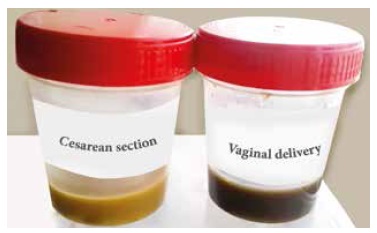
Meconium samples from those born by cesarean section were greenish-yellow, while meconium samples from those born vaginally were greenish-black (Figure 1). During the recollection of meconium, there were differences in the samples from two infants born by cesarean section, which corresponded to 3-days-old infants; the remaining samples corresponded to 1-day-old infants. Likewise, 28 of the neonates born by cesarean section were fed with artificial formula and two of them with maternal milk from the third day of life; while those born vaginally were exclusively breastfed.
Isolation of bacteria
In the pre-enrichment stage, two meconium samples from newborns by cesarean section formed gases that released unpleasant odors, said samples were then planted in MacConkey agar. After incubation, the presence of colonies of enterobacteria was confirmed.
In the plate growth test, 28 of 30 meconium samples from vaginal newborns showed growth of colonies of lactic acid bacteria (93.3%) compared to two of 30 meconium samples from those born by caesarean section (6.7%) (Figure 2). Likewise, the average number of colonies was higher in the meconium of those born vaginally (p<0.001) (Table 1). These colonies were observed under the microscope, and long thin bacilli and short bacilli were found, which correspond to characteristics of lactic acid bacteria (9). A total of 48 colonies were isolated and planted in modified MRS agar, and a better differentiation of the colonies according to size, color and border was observed (Figure 3).
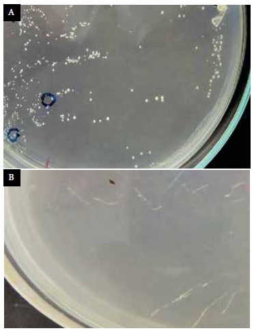
Figure 2 Growth of colonies on MRS agar. A) There is evidence of increased growth of acidic bacteria from vaginal newborns. B) Compared to those born by cesarean section.
Table 1 Count of lactic acid bacteria developed on MRS agar and acid pH resistance test, in vaginal newborns and by cesarean section.
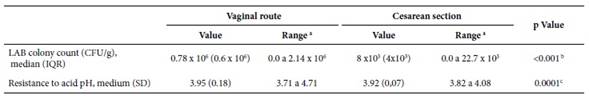
a Minimum - maximum; b Mann Whitney U-test; c Student’s t-test
IQR: interquartile range; SD: standard deviation; LAB: lactic acid bacteria, CFU: colony forming units
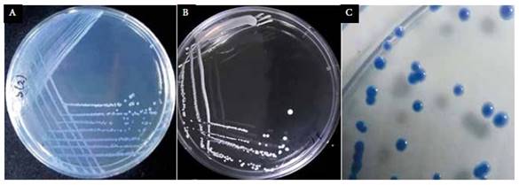
Figure 3 Isolation of colonies on MRS agar from the meconium of vaginal newborns. A) MRS agar modified with blue bromophenol to differentiate colonies of lactic acid bacteria. B) Agar without growth modified MRS. C) Modified MRS agar with isolated colonies of round shape characteristic of lactic acid bacteria
Tolerance to bile salts and acid pH
Forty-eight strains of lactic acid bacteria were isolated and subjected to a tolerance of >30% survival for 24 hours at an acidic pH of 3.0, and >20% survival at 0.3% bile salt concentration. A total of 33 (68.7%) surviving strains were obtained with greater resistance to acid than to bile salts, measured by absorbance.
DISCUSSION
It was shown that meconium from vaginal newborns had a higher presence of lactic acid bacteria than meconium from cesarean newborns, and little presence of anaerobic organisms.
The growth of acid-forming bacteria in samples of meconium from vaginal newborns is related to the largest quantity of lactobacilli in a woman’s vagina, which, during childbirth, is transmitted to and colonizes the mucous membrane of the digestive, respiratory, urogenital and skin tracts of the newborn 13. Intestinal colonization occurs especially by the genus Lactobacillus, which contributes to the formation of microbiota in neonates (14-15. Therefore, we consider that the increased growth of lactic acid bacteria in the meconium of vaginal newborns was due to the passing through the birth canal and the exclusive breastfeeding they received.
In contrast, meconium in newborns born by cesarean section showed a greater presence of enterobacteria, which would be directly related to the presence of the genera Clostridium, Staphylococcus, Propiobacterium and Corynebacterium in the digestive tract of these newborns 14-15. In addition, recent research indicates that those born by cesarean section would lack intestinal colonization, or that this would occur late, even up to one year of age 16. Therefore, the low growth of lactic acid bacteria in the meconium of newborns born by caesarean section (6.7%) would be related to the low consumption of breast milk on the third day of life, since the presence of lactic acid bacteria (Lactobacillus, Staphylococcus, Streptococcus and Bifidobacterium) has been reported in breast milk, which is transmitted to the newborn when it is breastfed 17.
The greenish-yellow color and the characteristic strong smell of feces from cesarean
section newborns, presumably from formula feeding, is different from that observed in vaginal infants who were fed primarily breast milk 13.
The characteristics described of the counted and selected white colonies in solid medium correspond to the description of lactic acid bacteria of the genus Lactobacillus (9. These bacteria, growing in MRS broth, showed rapid precipitation of white cells at the bottom of the tube, which occurs when growth ceases, giving rise to a soft and homogeneous sediment 18.
Of the total strains of lactic acid bacteria isolated, 68.7% showed resistance to gastrointestinal conditions during an exposure time of 24 hours measured by absorbance. This value is less than the 73.3% reported for Lactobacillus ramosus exposed for two hours 19, the difference could be related to the exposure time. It is likely that the surviving strains were Lactobacillus casei (ATCC 27139, FAGRO 98, FAGRO 98LP) and Lactobacillus rhamnosus (FAGRO 36.5), since their resistance to acid pH and high bile salt concentrations has been reported, even surviving in the gastrointestinal tract 20.
All the isolated lactic acid bacteria fermented the milk lactose and the average pH recorded was 3.95, which is within the range of Lactobacillus spp (12.
Among the study limitations, it should be mentioned that, the samples extraction was limited to two to three per week, for each group evaluated. The time interval between the collection of each sample depended on the defecation of the neonate, which influenced its transfer to the laboratory for the corresponding analyses. However, once the sample was collected, it was kept in adequate conditions to avoid contamination and deterioration.
Finally, it can be concluded that meconium from vaginal newborns showed a greater development of lactic acid bacteria with a greater presence of bacteria of the genus Lactobacillus compared to those born by cesarean section, in whom the presence of enterobacteria was evident.
Acknowledgements:
To Dr. Luz Veliz Sedano and Dr. Aida Cordero for their valuable contributions to the development of this research.
REFERENCES
1. Harmsen HJM, Wildeboer-Veloo ACM, Raangs GC, Wagendorp AA, Klein N, Bondels JG, et al. Analysis of intestinal flora development in breast-fed infants by using molecular identification and detection methods. J Pediatr Gastroenterol Nutr. 2000;30(1):61-7. [ Links ]
2. Jiménez EA. Fuentes de bacterias para la colonización del intestino del neonato: aplicación para el tratamiento de la mastitis lactacionales. [Tesis Doctoral]. Madrid: Universidad Complutense de Madrid; 2010 [citado el 20 de junio de 2017]. Disponible en https://eprints.ucm.es/10063/1/T31541.pdf. [ Links ]
3. Grönlund MM, Lehtonen OP, Ferola E, Kero P. Fecal microflora in healthy infants born by different methods of delivery: permanent changes in intestinal flora after cesarean delivery. J Pediatr Gastroenterol Nutr. 1999;28(1):19-25. [ Links ]
4. Mitsou EK, Kirtzalidou E, Oikonomou I, Liosis G, Kyriacou A. Fecal microflora of Greek healthy neonates. Anaerobe. 2008;14(2):94-101. doi: 10.1016/j.anaerobe.2007.11.002. [ Links ]
5. Marild K, Stephansson O, Montgomery S, Murray JA, Ludvigsson JF. Pregnancy outcome and risk of celiac disease in offspring: a nationwide case-control study. Gastroenterology. 2012;142:39-45. doi: 10.1053/j.gastro.2011.09.047. [ Links ]
6. Aumeunier A, Grela F, Ramadan A, Pham Van L, Bardel E, Gomez Alcala A, et al. Systemic Toll-like receptor stimulation suppresses experimental allergic asthma and autoimmune diabetes in NOD mice. PLoS ONE. 2010;5(7):e11484. doi: 10.1371/journal.pone.0011484. [ Links ]
7. Blustein J, Attina T, Liu M, Ryan AM, Cox LM, Blaser MJ, et al. Association of caesarean delivery with child adiposity from age 6 weeks to 15 years. Int J Obes (Lond). 2013;37(7):900-6. doi: 10.1038/ijo.2013.49. [ Links ]
8. Moreno R, Salas EJ, Pérez CI, Jiménez J. Evaluación del potencial probiótico de Lactobacilos aislado de heces de lactantes y leche materna. Rev Fac Med ULA. 2011;20(1):135-9. [ Links ]
9. Kandler O, Weiss G. Regular nonsporing Gram positives rods. En: Sneath PHA, Mair NS, Sharpe ME, Holt JG, editors. Bergey's Manual of Systematic Bacteriology. Vol 2. Baltimore: Maryland; 1986. pp. 1208-60. [ Links ]
10. Mejía-Rodríguez JA, Chacón-Rueda Z, Guerrero-Cárdenas B, Ottoniel-Rojas J, López-Corcuera G. Obtención de cepas de Lactobacillus: Caracterización in vitro como posible potencial probiótico. Rev Cientif. 2007;17(2):178-85. [ Links ]
11. Lara C, Burgos A. Potencial probiótico de cepas nativas para uso como aditivos en la alimentación avícola. Rev Colomb Biotecnol. 2012;14(1):31-40. [ Links ]
12. Sánchez L, Omura M, Lucas A, Pérez T, Llanes M, Luce C. Cepas de Lactobacillus spp. Con capacidades probióticas aisladas del tracto intestinal de terneros neonatos. Rev Salud Anim. 2015;37(2):94-104. [ Links ]
13. Gritz EC, Bhandari V. The Human Neonatal Gut Microbiome: A Brief Review. Front Pediatr. 2015;3:17. doi: 10.3389/fped.2015.00017. [ Links ]
14. Biasucci G, Rubini M, Riboni S, Morelli L, Bessi E, Retetangos C. Mode of delivery affects the bacterial community in the newborn gut. Early Hum Dev. 2010;86(1):13-5. doi: 10.1016/j.earlhumdev.2010.01.004. [ Links ]
15. Madan JC, Salari RC, Saxena D, Davidson L, O'Toole GA, Moore JH, et al. La colonización microbiana del intestino en neonatos prematuros predice la sepsis neonatal. Arch Dis Child Fetal and Neonatal Edition. 2012;97(6):F456-62. [ Links ]
16. Jakobsson HE, Abrahamsson TR, Jenmalm MC, Harris K, Quince C, Jernberg C. Decreased gut microbiota diversity, delayed Bacteroidetes colonisation and reduced Th1 responses in infants delivered by caesarean section. Gut. 2014;63(4):559-66. doi: 10.1136/gutjnl-2012-303249. [ Links ]
17. Solís G, De los Reyes-Gavilan CG, Fernández N, Margolles A, Gueimonde M. Establishment and development of lactic acid bacteria and bifidobacteria microbiota in breast-milk and the infant gut. Anaerobe. 2010;16(3):307-10. doi: 10.1016/j.anaerobe.2010.02.004. [ Links ]
18. Holt JG, Krieg NR, Sneath PHA, Staley JT, Williams ST. Bergey´s Manual of Determinative and Bacterology 9a ed. Washington: National Academy Press; 2001. [ Links ]
19. Rodríguez Gonzáles M. Aislamiento y selección de cepas del género Lactobacillus con capacidad probiótica e inmunomoduladora. [Tesis Doctoral]. Barcelona: Universidad de Barcelona; 2009 [citado 20 enero 2019]. Disponible en: https://www.tdx.cat/bitstream/handle/10803/3931/mrg1de1.pdf. [ Links ]
20. León Reissig MF. Evaluación in vitro de cepas de bacterias acidolácticas nativas con potencial probiótico. [Tesis de Licenciatura]. Montevideo: Universidad de la Republica; 2012. [citado 12 marzo 2019]. Disponible en: https://www.colibri.udelar.edu.uy/jspui/handle/20.500.12008/1432. [ Links ]
Funding sources: Self-fund research and financial support of Fundacion para el Desarrollo Agrario of Universidad Nacional Agraria La Molina.
Citation: Paitán-Anticona E, Sotelo-Méndez A, Bernuy Osorio N, Sumarriva-Bustinza L, Norabuena Sotelo A. Influence from the type of birth on the content of lactic acid bacteria in neonate meconium. Rev Peru Med Exp Salud Publica. 2020;37(1):93-98. Doi: https://doi.org/10.17843/rpmesp.2020.371.4251
Received: February 06, 2019; Accepted: January 08, 2020
 Este es un artículo publicado en acceso abierto bajo una licencia Creative Commons
