


Servicios Personalizados
Revista

Articulo
 texto en
  Inglés (pdf)
 Articulo en XML
 Referencias del artículo
 Enviar articulo por email
Indicadores
-
 Citado por SciELO
Links relacionados
-
 Similares en
SciELO
Similares en
SciELO
Compartir

 Permalink
Revista Peruana de Medicina Experimental y Salud Publica
versión impresa ISSN 1726-4634
Rev. perú. med. exp. salud publica vol.38 no.3 Lima jul./sep 2021 Epub 30-Sep-2021
http://dx.doi.org/10.17843/rpmesp.2021.383.6937
Original articles
Toxicity assessment of synthetic chalcones with antileishmanial potential in BALB/c mice
, Biologist http://orcid.org/0000-0002-6343-8785
, Bachelor’s Degree in Pharmacy and Biochemistry, Master’s Degree in Biochemistry and Molecular Biologyhttp://orcid.org/0000-0003-4349-3477
, Bachelor’s Degree in Pharmacy and Biochemistryhttp://orcid.org/0000-0002-7393-6147
, Chemist, PhD in Organic Chemistryhttp://orcid.org/0000-0002-1675-9105
, Biologist, Doctor of Sciencehttp://orcid.org/0000-0003-3837-2849
, Pharmaceutical Chemist, PhD in Pharmacochemistryhttp://orcid.org/0000-0003-3919-532X
, Biologist, PhD in Biomedical Scienceshttp://orcid.org/0000-0001-8516-006X
, Biologist, Master’s Degree in Biochemistry and Molecular Biologyhttp://orcid.org/0000-0003-1010-2353
1 Laboratorio Mixto Internacional Andino-Amazónico de Química de la Vida IRD-UPCH, Universidad Peruana Cayetano Heredia, Lima, Perú.
2 UMR 152 PHARMA-DEV, Université de Toulouse, IRD, Toulouse, Francia.
3 Laboratorio de Biología Celular y Molecular de Tripanosomátidos, Instituto de Medicina Tropical Alexander von Humboldt, Universidad Peruana Cayetano Heredia, Lima, Perú.
Objective:
To evaluate the toxicity of three synthetic chalcones administered intraperitoneally to BALB/c mice.
Materials and methods:
The median lethal dose (LD50) was estimated by Dixon’s Up-and-Down method. Subchronic toxicity of chalcones was evaluated at 20 and 40 mg/kg for 21 days. Behavioral, physiological, biochemical, and histological toxic effects were evaluated.
Results:
Chalcone 43 produced mucus in feces, visceral damage (liver) and alterations in organ coefficient (kidney, p = 0.037 and brain, p = 0.008) when compared to the control group. In addition, histological analysis showed that this chalcone produced edema, inflammation and necrosis in the evaluated organs, although there was no significant difference with the control. None of the biochemical parameters differed significantly between the treatment groups at 40 mg/kg dose and the control.
Conclusions:
The LD50 for all three chalcones was greater than 550 mg/kg of body weight. Chalcones 40 and 42 were found to be relatively non-toxic. Both can be considered safe for intraperitoneal application in BALB/c mice and, consequently, are potential candidates for use in the treatment of leishmaniasis.
Keywords: Synthetic Chalcones; Drug Development; BALB/c Mice; Subchronic Toxicity; Median Lethal Dose (source: MeSH NLM).
INTRODUCTION
Chalcones are abundant in edible plants, are part of the flavonoid family and have a wide spectrum of pharmacological activities 1 , 2. Regarding their antiparasitic properties, their activity against pathogenic protozoa of the Trypanosomatidae family is noteworthy, which includes the Trypanosoma genus (which causes Chagas disease and human African trypanosomiasis) and Leishmania (which causes leishmaniasis) 3 , 4. Several studies report the chemical synthesis and evaluation of natural chalcone analogues with the aim of developing compounds with improved selective antitrypanosomatid activity 3 - 5, including chalcone derivatives with promising arginase inhibitory activity in Leishmania infantum 6. These studies show the constant effort to search for new therapeutic alternatives against trypanosomiasis and leishmaniasis, which affect millions of people and animals worldwide ( 7, and for which the few available treatments have limitations 8.
In previous studies, with a new series of synthetic chalcones, our team evaluated in vitro activity against Leishmania species responsible for New World cutaneous and mucosal leishmaniasis. Nine chalcones (including 40, 42, and 43) showed biological activity and selectivity comparable to amphotericin B on both axenic amastigotes and intracellular amastigotes in macrophage infections, with selectivity indexes ranging from 10 to 80 9. Five chalcones were evaluated in vivo in BALB/c mice infected with Leishmania amazonensis (using the classical model of subcutaneous inoculation of the parasite in the plantar pad). Two of these compounds (42 and 43), administered intralesionally, showed a reduction in parasite load compared to untreated infected controls 9. For this evaluation, the compounds were administered at a dose of 5 mg/kg/day (a dose almost 7 times lower than the positive control with glucantime, the first-line reference drug, administered at 33 mg/kg/day). Based on these results and in order to guide the development of a potential new antileishmanial drug, we decided to evaluate the toxicity in in vivo experimental models taking into account the administration route of the compounds, dosage and treatment schedule, similar to that used in clinical treatment with reference drugs. This investigation aimed to determine the mean lethal dose and possible subchronic toxicity of three synthetic chalcones (40, 42 and 43) administered intraperitoneally in BALB/c mice.
KEY MESSAGES
Motivation for the study: Chalcones have a broad spectrum of antiparasitic activity, which is why they were evaluated as intralesional treatment against cutaneous leishmaniasis in a previous study. The toxicity of these compounds applied intraperitoneally in in vivo models has not yet been described.
Main findings: All three evaluated synthetic chalcones had a median lethal dose (LD50) greater than 550 mg/kg body weight, Chalcone 43 produced visceral damage and edema in internal organs unlike chalcones 40 and 42.
Implications: The relatively non-toxic chalcones 40 and 42 may be part of future therapeutic strategies against leishmaniasis.
MATERIALS AND METHODS
Design
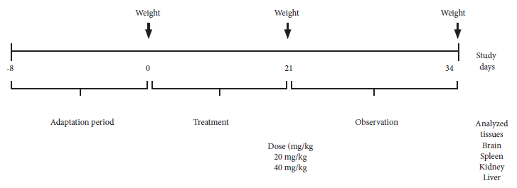
This was an experimental study that was carried out with the three chalcones (40, 42 and 43) previously identified by our team. For the determination of the LD50, female mice (n = 6) were used because they are more susceptible to the compounds according to the Organization for Economic Cooperation and Development (OECD) 10. The LD50 was estimated using the Up-and-Down method 11. To determine subchronic toxicity, animals were randomly distributed into eight groups (6 male mice per group). Two doses per chalcone were chosen according to the reviewed literature 4: 20 mg/kg (dose A) and 40 mg/kg (dose B) of animal weight. Each dose was administered according to the treatment schedule: nine administrations in 21 days (three per week) in a maximum volume of 100 µL intraperitoneally (IP). We included a control group, to which the solvents used in the compound dissolution were applied, and the Sham group, which did not receive any treatment. The experimental design for recording clinical data (body weight, organ coefficient, mortality, blood biochemistry and histopathology) is described in Figure 1.
Procedures
Animals
BALB/c mice (8 - 10 weeks old, 30 g weight and pathogen-free) were obtained from the National Institute of Health of Peru. The vivarium was maintained at 22 °C ± 2 °C and 60% ± 10% relative humidity with a 12-h light-dark cycle. The animals received food and water ad libitum.
Chemicals
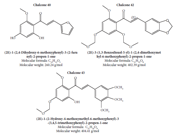
The de novo synthesis of chalcone 40 (C14H12O5) was carried out following the methodology described by Aponte et al. 3 from 2’,4’,6’-trihydroxyacetophenone, while chalcone 42 (C21H22O8) and chalcone 43 (C21H24O8) were synthesized by a similar route described by Du et al. 12.
The identity and purity of the compounds were verified by nuclear magnetic resonance (NMR) spectroscopy (Figure 2). The spectra were recorded at the NMR service of the Pontificia Universidad Católica del Perú. For IP application in mice, the compounds were dissolved in 5% dimethyl sulfoxide (DMSO) and 4% Tween 20 in 0.01 M phosphate-buffered saline (PBS).
Estimation of the LD 50 using the Dixon Up-and-Down Method
The first animal receives a dose below the best preliminary estimate of the oral LD50 11. If the animal survives, the second animal receives a higher dose. If the first animal dies or appears moribund, the second animal receives a lower dose. The recommended dose to use when there is no previous data is 175 mg/ kg body weight; the experiment started with this dose and then 550 mg/kg body weight was used. The animal was evaluated 48 hours after each dose. During this period (at 15 minutes, 30 minutes, 24 hours and 48 hours) we applied the modified Irwin test ( 13. The animals were euthanized six days after the administration of the doses, for macroscopic examination of the liver, spleen and kidney.
Subchronic toxicity study
Animals were examined individually at least once during the first 30 minutes after IP administration, periodically during the first 24 hours and then daily for 13 days after the last administration (until day 34), except when required to be removed from the study for animal welfare reasons or were found dead.
Body weight and organ coefficient
Animals were euthanized according to the euthanasia protocol of the Institutional Animal Use Ethics Committee (CIEA) of the Universidad Peruana Cayetano Heredia. The liver, spleen, kidneys and brain were removed and weighed. In addition, we included sections of intestine. We determined organ weight in relation to body weight (organ coefficient).
Determination of biochemical parameters
Before being euthanized, the animals were transferred to cages, without food for six hours, before blood sampling by cardiac puncture. Serum was obtained by centrifugation at 3000 g for 10 min. The measured parameters included two enzymes indicative of hepatocellular effects: aspartate aminotransferase (AST/GOT) and alanine aminotransferase (ALT/GPT); creatinine, C-reactive protein and glucose. These biochemical parameters were determined using commercial kits (Wiener lab., Argentina). The commercial kits do not require reference values for animals. The analysis was carried out comparing the treated groups with the control group.
Histopathology
Organs were embedded in paraffin; then, 4 µm sections were stained with hematoxylin-eosin using standard techniques. We analyzed each specimen by light microscopy in five fields at 20× magnification. The pathologist performing the histopathology was unaware of the identity of the specimens. The fields were selected randomly and continuously. A qualitative evaluation of the material was carried out 4, where: absent = 0, mild = +, moderate = ++ and severe = +++. The histopathological characteristics we recorded were: edema, inflammation and necrosis.
Statistical analysis
To determine the normal distribution of the data, we used the normal probability plot (QQ plot). Given this compliance, the comparison of the data (weight, organ coefficient and biochemical values) between the study groups was conducted with ANOVA. To identify the significance value between groups, we applied multiple comparison of means according to Tukey’s criterion. For comparison of histological findings (edema, inflammation and necrosis) between groups, we used Chi-square and Fisher’s tests. R program version 3.3.2 for Windows was used. We considered a significance level of 0.05.
RESULTS
LD50 according to Dixon’s Up-and-Down Method
One animal per dose per chalcone was used to determine the LD50. Oral administration of chalcones (40, 42 and 43) was initiated at a dose of 175 mg/kg body weight to one animal. During the first 48 hours of qualitative evaluations of behavior, neurological and autonomic characteristics, no lethality, convulsions, sedation, excitation, motor incoordination, aggressiveness, abnormal gait, jumping, abdominal writhing, piloerection or stereotypies (sniffing, chewing or head movements) were observed. The animals survived the observation period; therefore, we administered the next dose of 550 mg/kg body weight.
As with the previous dose, the treated animals (chalcone 40, 42 and 43) showed no evidence of alterations in behavior, neurological and autonomic characteristics during the six days of observation. The necropsy showed: chalcone 40, splenomegaly; chalcone 42, organs without alterations; chalcone 43, anatomical alterations such as visceral distress and edema (in intestine and liver), fibrin formation in liver, fusion of liver with diaphragm and formation of ligaments between the external walls of the intestine. The alterations due to chalcone 43 were observed in the animals that received both doses (175 mg/kg and 550 mg/kg body weight).
Subchronic toxicity study
After the second dose, all animals presented a brief lethargy (30 minutes) and abdominal pain. After this period, the animals resumed their regular activity. In addition, it was possible to observe changes in the behavior of the mice (aggressiveness) with chalcone 43 (20 mg/kg and 40 mg/kg) from the end of treatment (day 21) until the end of the experiment (day 34). Aggressiveness was not evident in the animals treated with chalcone 40 and 42 at the two doses administered until the end of the experiment. In all groups treated with chalcones, no apparent change in the hair or in the eyes were evident.
Animals in the group treated with chalcone 43 (40 mg/kg) presented mucus in the feces at 24 hours after the first dose was administered. This condition continued until day 34. On the other hand, in the groups treated with chalcone 42 and chalcone 40 (20 mg/kg and 40 mg/kg), mucus was observed between application 4 (day 8) and 6 (day 11). At the end of treatment, animals in the control group presented the same sign. No signs of colitis were evident in the mice with mucus in the feces. Figure 3 shows the results of the weight gained by the animals at the end of treatment compared to day 1 (start of treatment). The groups treated with chalcone 42 (40 mg/kg) or chalcone 43 (20 mg/kg and 40 mg/kg) did not show weight gain at day 21 compared to the control group (p > 0.05). However, there was a statistically significant difference in weight gain in groups 42 (40 mg/kg) (p = 0.037) and 43 (20 mg/kg) (p = 0.033) compared to the Sham group. Two weeks after the end of treatment (day 34), all treated groups showed modest weight gain, with the exception of groups 40 and 42, both at the 40 mg/kg dose.
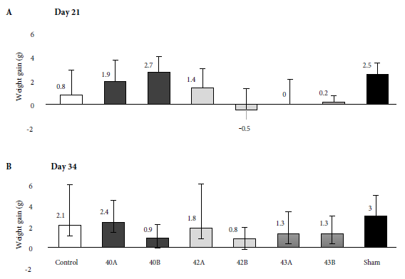
Figure 3 Weight gain of control animals versus those treated with synthetic chalcones 40, 42 and 43 (where A: 20 mg/kg and B: 40 mg/kg) at the end of treatment compared to day 1 (start of treatment). A: day 21. B: day 34. (n = 6 per group). Data are expressed as mean and standard deviation.
The organ coefficient results (Figure 4) showed that chalcones affected the internal organs we evaluated. Chalcone 42 at the dose of 20 mg/kg (42A) reduced the spleen coefficient (p = 0.034), while chalcone 43, at the dose of 40 mg/kg (43B), increased the kidney coefficient (p = 0.037) and the brain coefficient (p = 0.008). No significant differences were observed for the liver.
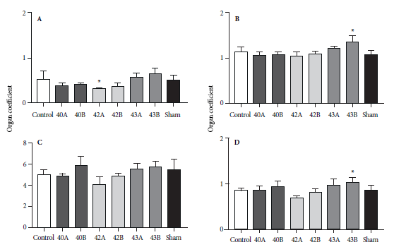
Figure 4 Organ coefficient from BALB/c mice treated with synthetic chalcones 40, 42 and 43 (where A: 20 mg/kg and B: 40 mg/kg). A: spleen, B: brain, C: liver, D: kidney. Control: chalcone diluent. Sham: untreated animals. Data are expressed as mean and standard deviation (6 per group). *p < 0.05 compared to control group, using ANOVA.
Creatinine decreased due to the treatment with chalcones 42 and 43, both administered at the dose of 20 mg/kg, compared to the control group (mean ± standard deviation: 2.20 ± 1.31; p = 0.026 and 2.50 ± 0.74; p = 0.033 vs. 16.30 ± 2.09 mg/L, respectively). No significant variations were found for GOT and GPT transaminase levels influenced by treatment (Figure 5). C-reactive protein was not detected in any sample.
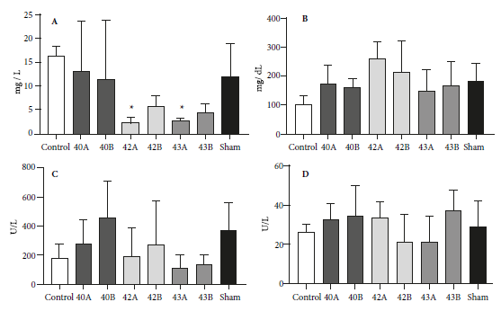
Figure 5 Analysis of biochemical parameters in serum of male BALB/c mice treated with synthetic chalcones 40, 42 and 43 (where A: 20 mg/kg and B: 40 mg/kg). Control: chalcone diluent. Sham: untreated animals. A: creatinine, B: glucose, C: GOT, D: GPT. Data are expressed as mean and standard deviation (6 per group). *p < 0.05 compared to control group, using ANOVA.
All groups had at least one mouse, with at least one organ histologically altered (mild/moderate level in the control and Sham groups; moderate/severe in the treated groups). Necrosis in the liver, spleen, kidney and brain was not evident in any group (including the control); neither was inflammation in the liver or spleen (Figure 6). It is noteworthy that histological alterations were severe in the intestines of treated mice (mainly with chalcone 43). Despite the histological alterations, there was no statistically significant difference (p > 0.05) between the chalcone-treated groups compared to the control and the Sham group.
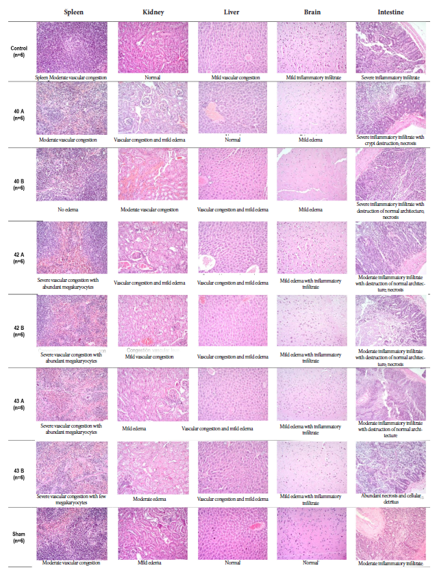
Figure 6 Representative images of histopathological evaluation of mouse vital organs from the subchronic toxicity study of synthetic chalcones. Representative sections of spleen, kidney, liver, brain and intestine stained with hematoxylin-eosin, taken from untreated (Sham), control (solvent), mice administered with 20 mg/kg synthetic chalcones (40A, 42A and 43A) or 40 mg/kg synthetic chalcones (40B, 42B and 43B). 200× magnification.
DISCUSSION
In this study, the subchronic in vivo toxicity of synthetic chalcones 40, 42 and 43 was evaluated by IP route following the reference treatment scheme for leishmaniasis; as well as the LD50, by oral route. The three chalcones, at a dose of 40 mg/kg, did not alter biochemical serum parameters. On the other hand, some of the anatomo-histopathological characteristics of main organs, organ coefficients and behavioral signs in the animals were altered, mainly in those treated with chalcone 43.
Few studies have reported the analysis of the in vivo toxicity of chalcones and their ADME process (absorption, distribution, metabolism and excretion) in the organism, despite their simple chemical structure and promising effects at the level of biological activity. These studies have mainly evaluated the biological activity in different pathological models 2 - 4 , 6 , 9 , 14 , 15. Based on the results of our study, we suggested to address ADME processes mainly with chalcones 40 and 42 in future studies.
Depending on the in vivo leishmaniasis model, the administration route, treatment schedule and biological effect may differ. De Mello et al. determined the non-toxicity of two synthetic chalcones (intralesionally and IP) up to a dose of 10 mg/kg body weight administered three times a week for four weeks and their antiparasitic activity against Leishmania braziliensis in golden hamsters 16. Aponte et al. observed high in vivo activity of chalcones 42 and 43 (administered intralesionally) in the murine model infected by Leishmania amazonensis in the plantar pad 9. Considering the above, the importance of selecting the best administration route is validated when the compounds are designed to be applied as a treatment, in such a way that bioavailability is favored and their biological activity is maximized.
Intraperitoneal administration of drugs is widely used in rodents because it improves the absorption speed compared to the intramuscular and subcutaneous routes 17 , 18. The main limitation of the IP route is the effect of initial metabolism; however, low molecular weight drugs (such as chalcones) administered by the IP route are considered to result in lower systemic exposure 17. Other limitations include the sterility and irritancy they may generate 17. Irritant compounds can cause ileus and peritoneal inflammation and this could favor the development of adhesions, which were evident in the necropsy of animals treated with chalcone 43. Although the route of administration differs (oral route, 1000 mg/kg/day) from that used in this study, it has been reported that other chalcones can affect some physiological parameters, such as kidney weight, altering the organ coefficient, and generate minimal macroscopic changes in the liver 19, as recorded in the animals of the chalcone 43B group (40 mg/kg) in this study. In contrast, other authors have not reported histological changes in the kidney or liver, nor the alteration of biochemical parameters generated by chalcones administered intralesionally 15.
The absorption of drugs applied in the peritoneal cavity is caused by diffusion to the surrounding tissues (including the intestine) 17 , 18. In this study, we propose that IP administration and intestinal diffusion of chalcones may cause an increased amount of mucus in the feces, which acts as a biological barrier, mainly in animals treated with chalcone 43, this being confirmed at the histopathological level. There is limited information regarding the toxicological relevance of the interaction between mucus in feces and xenobiotic compounds for the host. Studies indicate that the amount of intestinal mucus can increase or decrease depending on the administered chemical compound or by diet 20 , 21. It is still poorly understood how environmental chemicals (pesticides, among others) can interact with intestinal mucus and the “mucophilic” bacteria of the microbiota 20.
It is to be expected that, despite environmental factors, organ coefficients for the same mouse strain will be similar. The control group organ coefficients determined in this study for the BALB/c strain are similar to those previously reported by Shi et al. for their control group 22. Commonly, abnormal body weight alterations in toxicity studies are considered critical indicators of adverse effects. The administered compounds and abdominal pain may influence the appetite of the animals and consequently the weight. However, in our study there was no significant difference in weight gained between the control and treated groups at the end of treatment (day 21). The weight gain recorded in our study is similar to that reported in mice by Zhao et al. 23 and Shi et al. 22.
The determination of subchronic toxicity in this study evidenced behavioral changes in animals treated with chalcone 43 (40 mg/kg). Although behavioral alterations (nervous system) due to chalcone derivatives have been reported previously 24; in our study, the aggressiveness of the animals was not validated by the histopathological analyses conducted. Considering that laboratory conditions, such as feeding, light exposure schedules, food and water availability, vary among institutions, as well as the methods used in the quantification of biochemical parameters, the generalization of reference values becomes complex. Mazzaccara et al. suggest using sex- and age-dependent hematological parameters for each mouse strain, as these vary from strain to strain 25. Although there are papers in the literature that discuss hematological and biochemical parameters for healthy BALB/c mice, few address the evaluation of these parameters in pathological situations 26; moreover, these have been obtained using different methodologies. Literature suggests to compare biochemical parameters between treated and untreated groups. Considering the approaches for obtaining reference values of hematological parameters in male BALB/c mice, the values of GPT and GOT found in this study are comparable to those established by Lima et al. 27 and Fernandes et al. 28. However, the values reported for creatinine in both studies are not comparable. The creatinine results in our study are below those reported by Lima et al. (0.58 ± 0.24 mg/dL) 27. On the other hand, Silva-Santana et al. reported undetectable reference values for creatinine (<0.2 mg/dL), determined with the automatic spectrophotometer VITROS® 350 (Ortho Clinical Diagnostics) 29. For a more sensitive screening, other parameters (alkaline phosphatase, triglycerides, blood urea nitrogen, among others) should be included to complement the detection of alterations in the animals’ metabolism.
According to Dixon’s method, trial continuation with doses higher than 550 mg/kg body weight is recommended. Given the limited availability of the synthetic compounds in this study, this was not possible. It should be noted that the maximum administered dose of the chalcones (550 mg/kg body weight, orally) was 100 times higher than the dose that proved to be effective (5 mg/kg/day, 8 inoculations intralesionally) in significantly reducing the parasite load in the mouse model infected by L. amazonensis in the plantar pad 9.
Using the chemical property predictor software admetSARTM version 2.0 30, the oral LD50 could be determined in silico in rats: chalcone 40 (5998.06 g/kg), chalcone 42 (10 986.05 g/kg) and chalcone 43 (9445.40 g/kg). In silico oral LD50 was determined in rats using the chemical property predictor software admetSARTM version 2.0 30: chalcone 40 (5998.06 g/kg), chalcone 42 (10 986.05 g/kg) and chalcone 43 (9445.40 g/kg).
In conclusion, the overall findings indicate that the LD50 for all three chalcones is greater than 550 mg/kg body weight. Chalcones 40 and 42 are relatively non-toxic and can therefore be considered for safe use in BALB/c mice. We consider it relevant to extend toxicity screening to chalcone analogues that show antileishmanial activity and are lead compounds candidates to guide the development of new drugs for leishmaniasis. Such studies should take into account the evaluation of toxicity, the administration route and the ADME process in in vivo models. This will contribute to the efforts of the scientific community 4 , 6 , 9 , 16 towards the validation the biological activity of chalcones in murine models of cutaneous and visceral leishmaniasis.
Acknowledgments:
The authors thank Luis Cano Ayestas, M.D., Ph.D., a specialist in anatomic pathology, for the histopathologic analysis.
REFERENCES
1. Nowakowska Z. A review of anti-infective and anti-inflammatory chalcones. Eur J Med Chem. 2007;42(2):125-37. doi: 10.1016/j.ejmech.2006.09.019. [ Links ]
2. Li W, Xu F, Shuai W, Sun H, Yao H, Ma C, et al. Discovery of Novel Quinoline-Chalcone Derivatives as Potent Antitumor Agents with Microtubule Polymerization Inhibitory Activity. J Med Chem. 2019;62(2):993-1013. doi: 10.1021/acs.jmedchem.8b01755. [ Links ]
3. Aponte JC, Verástegui M, Málaga E, Zimic M, Quiliano M, Vaisberg AJ, et al. Synthesis, cytotoxicity, and anti-Trypanosoma cruzi activity of new chalcones. J Med Chem. 2008;51(19):6230-4. doi: 10.1021/jm800812k. [ Links ]
4. de Mello TF, Bitencourt HR, Pedroso RB, Aristides SM, Lonardoni MV, Silveira TG. Leishmanicidal activity of synthetic chalcones in Leishmania (Viannia) braziliensis. Exp Parasitol. 2014;136:27-34. doi: 10.1016/j.exppara.2013.11.003. [ Links ]
5. Boeck P, Bandeira Falcão CA, Leal PC, Yunes RA, Filho VC, Torres-Santos EC, et al. Synthesis of chalcone analogues with increased antileishmanial activity. Bioorg Med Chem. 2006;14(5):1538-45. doi: 10.1016/j.bmc.2005.10.005. [ Links ]
6. García AR, Oliveira DMP, Jesus JB, Souza AMT, Sodero ACR, Vermelho AB, et al. Identification of Chalcone Derivatives as Inhibitors of Leishmania infantum Arginase and Promising Antileishmanial Agents. Front Chem. 2021;8:624678. doi: 10.3389/fchem.2020.624678. [ Links ]
7. Van Voorhis WC, Adams JH, Adelfio R, Ahyong V, Akabas MH, Alano P, et al. Open Source Drug Discovery with the Malaria Box Compound Collection for Neglected Diseases and Beyond. PLoS Pathog. 2016;12(7):e1005763. doi: 10.1371/journal.ppat.1005763. [ Links ]
8. Ghorbani M, Farhoudi R. Leishmaniasis in humans: drug or vaccine therapy?. Drug Des Devel Ther. 2017;12:25-40. doi: 10.2147/DDDT.S146521. [ Links ]
9. Aponte JC, Castillo D, Estevez Y, Gonzalez G, Arevalo J, Hammond GB, et al. In vitro and in vivo anti-Leishmania activity of polysubstituted synthetic chalcones. Bioorg Med Chem Lett. 2010;20(1):100-3. doi: 10.1016/j.bmcl.2009.11.033. [ Links ]
10. Organisation for Economic Cooperation and Development. Test No. 425: Acute Oral Toxicity: Up-and-Down Procedure, OECD Guidelines for the Testing of Chemicals, Section 4. París: OECD Publishing; 2008. doi: 10.1787/9789264071049-en. [ Links ]
11. Dixon WJ. The Up-and-Down method for small samples. J Am Stat Assoc. 1965;60(312):967-78. Disponible en: https://doi.org/10.2307/2283398. [ Links ]
12. Du Z, She X, Ma J, Wang Z, Wu H, Li Y, et al. A facile synthetic route to two chalcones. Journal of Chemical Research. 2004;2004(1):45-6. doi: 10.3184/030823404323000756. [ Links ]
13. Roux S, Sablé E, Porsolt RD. Primary observation (Irwin) test in rodents for assessing acute toxicity of a test agent and its effects on behavior and physiological function. Curr Protoc Pharmacol. 2005;Chapter 10:Unit 10.10. doi: 10.1002/0471141755.ph1010s27. [ Links ]
14. Staurengo-Ferrari L, Ruiz-Miyazawa KW, Pinho-Ribeiro FA, Fattori V, Zaninelli TH, Badaro-Garcia S, et al. Trans-Chalcone Attenuates Pain and Inflammation in Experimental Acute Gout Arthritis in Mice. Front Pharmacol. 2018;9:1123. doi: 10.3389/fphar.2018.01123. [ Links ]
15. Dhiyaaldeen SM, Amin ZA, Darvish PH, Mustafa IF, Jamil MM, Rouhollahi E, et al. Protective effects of (1-(4-hydroxy-phenyl)-3-m-tolyl-propenone chalcone in indomethacin-induced gastric erosive damage in rats. BMC Vet Res. 2014;10:961. doi: 10.1186/s12917-014-0303-7. [ Links ]
16. de Mello TF, Cardoso BM, Lopes SN, Bitencourt HR, Voltarelli EM, Hernandes L, et al. Activity of synthetic chalcones in hamsters experimentally infected with Leishmania (Viannia) braziliensis. Parasitol Res. 2015;114(10):3587-600. doi: 10.1007/s00436-015-4581-1. [ Links ]
17. Al Shoyaib A, Archie SR, Karamyan VT. Intraperitoneal Route of Drug Administration: Should it Be Used in Experimental Animal Studies? Pharm Res. 2019;37(1):12. doi: 10.1007/s11095-019-2745-x. [ Links ]
18. Intraperitoneal Drug Administration. En: Techniques in the Behavioral and Neural Sciences [Internet]. Elsevier; 1994 [citado el 23 de junio de 2021]; 12:46-58. Disponible en: https://linkinghub.elsevier.com/retrieve/pii/B9780444818713500102. [ Links ]
19. Maronpot RR. Toxicological assessment of Ashitaba Chalcone. Food Chem Toxicol. 2015;77:111-9. doi: 10.1016/j.fct.2014.12.021. [ Links ]
20. Gillois K, Lévêque M, Théodorou V, Robert H, Mercier-Bonin M. Mucus: An Underestimated Gut Target for Environmental Pollutants and Food Additives. Microorganisms. 2018;6(2):53. doi: 10.3390/microorganisms6020053. [ Links ]
21. Chassaing B, Koren O, Goodrich JK, Poole AC, Srinivasan S, Ley RE, et al. Dietary emulsifiers impact the mouse gut microbiota promoting colitis and metabolic syndrome. Nature. 2015;519(7541):92-6. doi: 10.1038/nature14232. [ Links ]
22. Shi JP, Li SX, Ma ZL, Gao AL, Song YJ, Zhang H. Acute and sub-chronic toxicity of tetrandrine in intravenously exposed female BALB/c mice. Chin J Integr Med. 2016;22(12):925-931. doi: 10.1007/s11655-015-2303-2. [ Links ]
23. Zhao Q, Yang M, Deng Y, Yu H, Wang L, Teng F, et al. The Safety Evaluation of Salvianolic Acid B and Ginsenoside Rg1 Combination on Mice. Int J Mol Sci. 2015;16(12):29345-56. doi: 10.3390/ijms161226176. [ Links ]
24. Kwak J, Kim MJ, Choi KC, Choi HK, Jun W, Park HJ, et al. The chalcone derivative Chana 1 protects against amyloid ß peptide-induced oxidative stress and cognitive impairment. Int J Mol Med. 2012;30(1):193-8. doi: 10.3892/ijmm.2012.984. [ Links ]
25. Mazzaccara C, Labruna G, Cito G, Scarfò M, De Felice M, Pastore L, et al. Age-Related Reference Intervals of the Main Biochemical and Hematological Parameters in C57BL/6J, 129SV/EV and C3H/HeJ Mouse Strains. PLoS One. 2008;3(11):e3772. doi: 10.1371/journal.pone.0003772. [ Links ]
26. Barbosa BDS, Praxedes ÉA, Lima MA, Pimentel MML, Santos FA, Brito PD, et al. Haematological and Biochemical Profile of Balb-c Mice. Acta Sci Vet. 2017;45(1):5. doi: 10.22456/1679-9216.80473. [ Links ]
27. Lima CM, Lima AK, Melo MGD, Dória GAA, Leite BLS, Serafini MR, et al. Valores de referência hematológicos e bioquímicos de ratos (Rattus novergicus linhagem Wistar) provenientes do biotério da Universidade Tiradentes. Sci Plena. 2014;10(034601):1-9. Disponible en: https://www.scientiaplena.org.br/sp/article/view/1784/948. [ Links ]
28. Fernandes DP, Pimentel MML, Santos FAD, Praxedes ÉA, Brito PD, Lima MA, et al. Hematological and biochemical profile of BALB/c nude and C57BL/6 SCID female mice after ovarian xenograft. An Acad Bras Cienc. 2018;90(4):3941-3948. doi: 10.1590/0001-3765201820180586. [ Links ]
29. Silva-Santana G, Bax JC, Fernandes DCS, Bacellar DTL, Hooper C, Dias AASO, et al. Clinical hematological and biochemical parameters in Swiss, BALB/c, C57BL/6 and B6D2F1 Mus musculus. Animal Model Exp Med. 2020;3(4):304-315. doi: 10.1002/ame2.12139. [ Links ]
30. Yang H, Lou C, Sun L, Li J, Cai Y, Wang Z, et al. admetSAR 2.0: web-service for prediction and optimization of chemical ADMET properties. Bioinformatics. 2019;35(6):1067-1069. doi: 10.1093/bioinformatics/bty707. [ Links ]
Funding: Fondo Nacional de Desarrollo Científico, Tecnológico y de Innovación Tecnológica del Consejo Nacional de Ciencia Tecnología e Innovación Tecnológica (CONCYTEC) (Convenio C-112- 2015 FONDECYT).
Received: December 18, 2020; Accepted: August 04, 2021
 Este es un artículo publicado en acceso abierto bajo una licencia Creative Commons
