


Servicios Personalizados
Revista

Articulo
 Inglés (pdf)
 Articulo en XML
 Referencias del artículo
 Enviar articulo por email
Indicadores
-
 Citado por SciELO
Links relacionados
-
 Similares en
SciELO
Similares en
SciELO  uBio
uBio
Compartir

 Permalink
Revista Peruana de Biología
versión On-line ISSN 1727-9933
Rev. peru biol. vol.25 no.4 Lima oct. 2018
http://dx.doi.org/10.15381/rpb.v25i4.15538
NOTA CIENTÍFICA
Description of the female of Pintomyia (Pifanomyia) deorsa (Diptera, Psychodidae, Phlebotominae)
Descripción de la hembra de Pintomyia (Pifanomyia) deorsa (Diptera, Psychodidae, Phlebotominae)
Eunice A. B. Galati 1, Gian C. Aramburu-Quispe 2, Carmen D. Rado-Covarrubias 2, Flor K. Toccas-Palomino 2, Elsa G. Aguilar-Ancori 2, Maritza M. Quispe-Florez 2, Rosa Pacheco 2, Elena Ogusuku 3, Marcia Bicudo de Paula 1, J. Enrique Perez 4*
1 Departamento de Epidemiologia, Faculdade de Saúde Pública, Universidade de São Paulo Av. Dr. Arnaldo 715, 01246-904 São Paulo, SP, Brasil.
2 Instituto Universitario de Enfermedades Tropicales y Biomedicina del Cusco, Universidad Nacional de San Antonio Abad del Cusco. Av. de la Cultura. 773, Cusco, Perú.
3 Dirección General de Salud Ambiental, Ministerio de Salud, Lima, Perú
4 Instituto de Medicina Tropical Alexander von Humboldt, Universidad Peruana Cayetano Heredia, Lima, Perú
*Autor para correspondencia
Email Eunice A. B. Galati: egalati@usp.br
Email Gian C. Aramburu-Quispe: caq-01@hotmail.com
Email Carmen D. Rado-Covarrubias: dirc82@gmail.com
Email Flor K. Toccas-Palomino: flor_13_8@hotmail.com
Email Elsa G. Aguilar-Ancori: egaa3@hotmail.com
Email Maritza M. Quispe-Florez: maritzamerc@yahoo.com
Email Rosa Pacheco: rpachec2@yahoo.com
Email Elena Ogusuku: elenaoa2013@gmail.com
Email Marcia Bicudo de Paula: bicudo@usp.br
Email J.Enrique Perez: kikelutz@yahoo.com
Abstract
Pintomyia (Pifanomyia) deorsa (Pérez, Ogusuku, Monje & Young, 1991) was described on the basis of a single male; the female is being described here from specimens collected in Ollantaytambo, Cusco, Peru. Diagnoses for the Pintomyia genus, Pifanomyia subgenus, Verrucarum series and both sexes of Pi. deorsa are presented, as well as an identification key to distinguish the females of the Verrucarum series.
Keywords: Bartonelosis; leishmaniasis; Vectors; Pintomyia; Andes.
Resumen
Pintomyia (Pifanomyia) deorsa (Pérez, Ogusuku, Monje & Young, 1991) fue descrita en base a un solo espécimen macho; la hembra es descrita aquí a partir de especímenes colectados en Ollantaytambo, Cusco, Perú. El diagnóstico para en género Pintomyia, el subgénero Pifanomyia, la série Verrucarum y ambos sexos de Pi. deorsa son presentados, así como claves para la identificación y separación de las hembras de la serie Verrucarum.
Palabras clave: Bartonelosis; leishmaniasis; Vectores; Pintomyia ; Andes.
Introduction
Phlebotominae are small dipterans with nocturnal activity, the females are mainly blood feeders, they take a bloodmeal through a biting to their preferred hosts, many times including human beings; several groups of pathogens take part of this situation, resulting in life cycles for the vector, and for the pathogens (Zorrilla et al. 2017). In Peru, nearly 200 species of Phlebotominae were reported; the fauna of the Western slopes and Interandean valleys of the Andean Cordillera includes at least four species (Perez et al. 1993), and the fauna of the Eastern slopes from 2000 down to 300 m of altitude is approximately 30 species (Perez et al. 1991). In Peru the Phlebotominae are known to transmit Leishmania spp. and Bartonella bacilliformis (Forattini 1973). Helcocyrtomyia peruensis is the most common species in the highlands of the Cusco region up to 3500 m of altitude (Grajeda et al. 2004).
Pintomyia deorsa has been described by Pérez et al. (1991) who included it in the Lutzomyia genus, Verrucarum group in accordance with Theodor’s classification (1965). Its description was made on the basis of a male collected from a rock crevice in Abancay outskirts, Apurimac Department, Peru, at 2380 m of altitude in October 1985.
The species, according to Galati’s classification (2003a) was included in the Verrucarum series, one of the seven of the subgenus Pifanomyia Ortiz & Scorza, 1963, which, in its turn, is one of the two subgenera of the genus Pintomyia Costa Lima, 1932 of the Lutzomyiina subtribe.
Despite of several collections undertaken in the region of the type-locality of Pintomyia (Pifanomyia) deorsa, with several specimens of Lutzomyia battistinii and some of Lutzomyia pescei being captured, no further specimens of Pi. deorsa have been collected there (Personal observation J. E. Pérez).
More recently two males of Pi. deorsa, together with some undescribed females, were collected with CDC light traps in October 2014 from Ollanta (2900-3200 m), a locality in Ollantaytambo, Urubamba Province, Cusco, Peru.
Other similar females were collected in the outskirts of Ollantaytambo city, Urubamba, Cuzco, (13°14’57.18"S; 72°15’ 36.37"W, 2916 m), with Shannon traps , extradomicile, using protected human beings as bait, during October 2015 and November 2016, Perez & Toccas colls.
This study presents a first description of the female of Pi. deorsa and some characteristics not mentioned in the original description but important for the characterization of the male of the group to which this insect belongs.
Material and methods
The specimens collected after clarification, according to Forattini (1973), were mounted on microscope slides in NC medium (Cerqueira 1943). The morphological characters were measured using the Zen version 2012, with images taken with an AxioCam camera model "105 color" (Carl Zeiss® MicroImaging GmbH, Jena, TH, DE) coupled to a light microscope. The drawings were made in pencil using an Olympus® microscope with the aid of an Olympus camera lucida. All measurements are given as averages, followed by standard deviation in micrometers, as well as the number of specimens measured.
The nomenclature adopted is from Galati (2003) and the characters are described in accordance with Galati et al. (2017). The abbreviation of the species name follows Marcondes (2007).
Material examined: A total of 37 specimens of Pi. deorsa collected during several trips to Ollantaytambo District, Urubamba Province, Cusco Department, Peru. The localities sampled were Ollanta (2 males and 35 females), and Pallata (1 female):
Ollanta S 13°14.57´18", W 72°15’36.38", 2918 m, 2 males (Shannon trap-peridomicile), October 17, 2014, Toccas and Aramburu colls., 1 female (CDC intradomicile) S 13°14’59.449", W 72° 15’39.936", October 20, 2014, Toccas and Aramburu colls., 2919 m, 3 females (CDC extradomicile), S 13°14’59.726", W 72°15’40.962", October 19, 2014, Cha-con & Farfan colls., 8 females (Shannon trap, 18-20 hours), August 30, 2015, Aramburu & Toccas colls., 13°14’57.18"S; 72°15’ 36.37"W, 2916 m, 1 female (CDC peridomicile) S 13°14’59.726", W 72°15’40.962", October 18, 2014, Toccas & Aramburu colls. 9 females (Shannon trap 18-21 hours), 2963 m, S 13°14’58.569", W 72°15’11.102", October 12, 2014, Chacon & Farfan colls., 4 females (Shannon trap: 18-20 hours) October 24, 2015, Perez & Toccas colls., 8 females (Shannon trap 18-21 hours), November 16, 2016, Perez & Toccas colls.
Pallata S 13°14’13.7", W 72° 13’26.5", 1 female (CDC extradomicile), October 19, 2014, Chacon & Farfan colls.,3176 m.
The material has been deposited in the entomological collection of the Faculdade de Saúde Pública da Universidade de São Paulo (FSP/USP), and the Instituto Universitario de Enfermedades Tropicales y Biomedicina del Cusco.
Results
Pintomyia
Diagnosis.- Both sexes: FI with the external ascoid implanted more apically than the internal one, 3rd palpal segment with the Newstead’s sensilla distributed in its middle or more apically, palpi with the 2nd segment as long as or longer than the 4th, ventrocervical sensilla present. Absence of postalar setae and the suture between katepisternum and metepimerum. Male with tergal papillae in at least one of the segments II to VII. Genitalia. Gonocoxite with a basal tuft of bristles and gonostyle with four well-developed spines with the presence of a preapical setiform spine and paramere simple, without protuberance in the pre-apical region of the ventral margin, epandrial lobe with rounded apex and narrower than the gonocoxite. The females present maxillary lacinia with one longitudinal row of external teeth. Cibarium with four posterior teeth and the anterior teeth arranged in one or two transversal rows. The spermathecae are predominantly vesiculous and the common spermathecal ducts long, reaching at least the middle of the stem of the genital furca.
Females, 8th tergite, generally, with setae.
Pintomyia (Pifanomyia)
Diagnosis.- Both sexes without spines in the ventral margin of the femur. Females with predominantly vesiculous spermathecae, but with an apical ring, individual spermathecal ducts not sclerotized.
Verrucarum series
Diagnosis.- Both sexes. Small eyes that in the frontal view present a smaller width than the interocular distance. Presence of papilla in FIII and absence of papillae in FXI. Male. Gonostyle with four spines and pre-apical spiniform seta. Gonocoxite. inner basal face with sclerotized longitudinal band, basal tuft of setae and generally other setae in the middle-apical region. Female. Apical ring of the spermathecae not collar-shaped.
Pintomyia (Pifanomyia) deorsa (Pérez, Ogusuku, Monje & Young, 1991)
Diagnosis.- Both sexes. Thoracic coloration: mesonotum and metanotum brown, pronotum and post-notum light brown, paratergite and pleura pale but pale brown at the base of the katepimeron, katepisternum and coxae. Head: antennal formula: FI-FXIII 2, FIV 0; Newstead’s sensilla absent from the 2nd palpal segment and a few grouped in the middle of the 3rd palpal segment. Antennae with short ascoids, not reaching the middle of the article in FII. Presence of simple setae in all flagellomeres.
Male.- Palpal formula: 1.4.3.2.5. Abdomen: presence of tergal papillae from 5th to 7th tergites. Genitalia: Gonocoxite: basal tuft with 10-12 setae; epandrial lobe longer than the gonocoxite; apex of the parameral sheath shaped like the neck and head of a goose. Gonostyle with the upper external spine implanted in its apical third and the lower external spine and the internal one in its middle; Aedeagal ducts/sperm pump ratio ca. 3.6:1.0
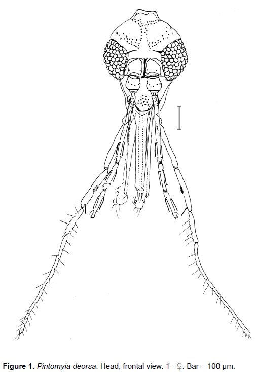
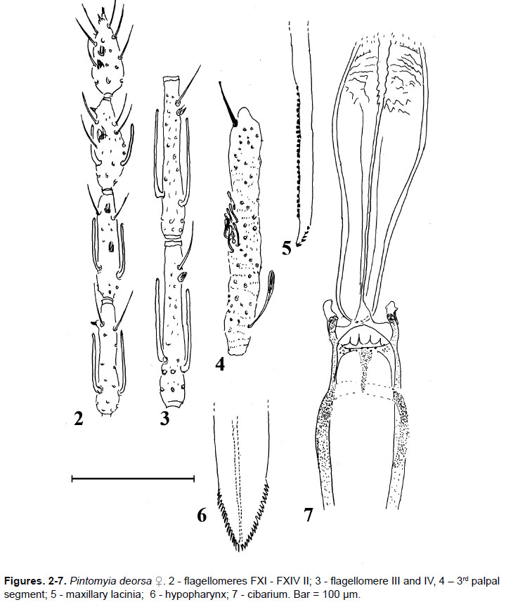
Female.-Total length: 3,084; 206 (n= 15). Head (Fig 1) 437.8; 18.8 (n = 23) long, 363.7; 24.4 (n = 20) wide, ratio between length/width 1.21:1.0; 0.07 (n = 20). Clypeus 150.3; 7.9 (n = 23) long; ratio clypeus length/head length 0.34:1.0; 0.01 (n = 20). Eye 191.1 ± 10.6 (n = 19) long; eye length/head length 0.44:1.0; 0.02 (n = 19). Eye 123.8; 13.9 (n = 19) wide. Interocular distance 143.1; 8.7 (n = 19). Labrum-epipharynx (LE) 396.9; 17.1 (n = 20) ratio: LE/head length 0.90:1.0 ± 0.04 (n = 22). Antenna: flagellomere lengths: FI 319.8; 16.0 (n = 11); FII 129.2; 5.6 (n = 11); FIII 131.3; 5.4 (n = 11); FXIII 70.6; 5.7 (n = 8; FXIV 61.8; 2.2 (n = 7). FI has its external ascoid implanted slightly more apically than the internal one; FII has the ascoid’s apex approaching the papilla level (Fig. 3); FIII with papilla (Fig. 3); FXI without papilla (Fig. 2) ratios: FI/head length 0.74:1.0; 0.03 (n = 10); FI/LE ratio 0.82:1.0; 0.05 (n = 10). Lengths of palpal segments: 1st 66.5; 8.4 (n = 24); 2nd 200.5; 14.8 (n = 24); 3rd 200.5; 8.9 (n = 21); 4th 126.3; 14.2 (n = 21); 5th 428.5; 41.3 (n = 21). Palpal formula 1.4.2.3.5 (n = 15) or 1.4 (2.3) (n =1) or 1.4.3.2.5 (n = 5); 2nd palpal segment without Newstead’s sensilla; 3rd palpal segment with ca. 10 Newstead’s sensilla situated in its middle (Fig. 4). Cibarium (Fig. 7) with two pairs of equidistant and needle-like horizontal posterior teeth; 6-10 (n = 20) anterior teeth very small, vertically positioned in relation to the lumen, arranged in one irregular transversal row, and also some others laterally grouped; the sclerotized area is clearly evident and funnel-shaped; sclerotized arch incomplete. The pharynx has atrophied spines in its apical quarter. Hypopharynx (Fig. 6) with 17-19 (n = 20) well-delimited short teeth situated apicolaterally. Lacinia of the maxilla (Fig. 5) presents 4-7 (n = 20) very short external teeth arranged in a single row, and 20-25 (n = 20) internal teeth. Labial sutures form a fork.
Ventrocervical sensilla present.
Thorax.- mesonotum 627.6; 39.1 (n = 23) long. Presence of 1-4 (n = 19) proepimeral setae and 4-14 (n = 26) upper anepisternal setae; setae on the anterior region of the katepisternum absent. Wing (Fig. 8) 2576.4; 115.2 (n = 20) long, 769.4; 49.9 (n = 20) wide; R5 1677.2; 63.4 (n = 20); alpha 653.8; 46.9 (n = 20); beta 217.0; 9.3 (n = 20); gamma 426.4; 27.9 (n = 20); delta 210.8; 45.9 (n = 20) ; pi 78.8; 19.5 (n = 20); length/width ratio: 3.4:1.0; 011 (n = 20); delta/alpha ratio: 0.3:1.0; 0.05 (n = 20). Legs, respectively anterior, median and posterior: femur 844.8; 59.2 (n = 11), 844.9;41.6 (n = 11), 910.2; 42.5 (n = 16); tibia 920.8;49.8 (n = 11), 1108.9; 51.4 (n = 11), 1388.9; 69.7 (n =16); tarsomere I 575.4; 19.2 (n = 11), 672.0;38.9 (n = 11), 823.6;51.2 (n = 15) and tarsomeres II+III+IV+V 760.5;41.2 (n = 9), 814.9; 43.0 (n = 8), 932.6;82.9 (n = 14).
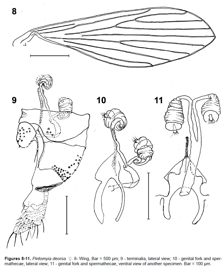
Abdomen.- 1,788; 189.1 (n = 22) long. Tergite VIII (Fig. 9) with 2 (n = 1), 3 (n = 1), 4 (n = 6), 5 (n = 7), 6(n = 8), 7(n = 3) or 8 (n = 1) setae on either side. Spermathecae length (Figs. 10, 11) - the measurements of their lengths are not shown because they were contracted, and 37.9; 3.5 (n = 11) of maximum width, with superficial striations and an apical ring. The transition to the individual sperm ducts is clear. These ducts are smooth, membranous, measuring 127.3; 32.2 (n = 11) in length by 7.2; 1.2 (n = 11) at maximum width; common sperm duct is also smooth and membranous, 99.6; 24.1 (n = 11) long by 11.8; 3.7 (n = 1) wide. Cercus 136.9; 14.9 (n = 19) long, with oblong apex.
The added male characteristics are presence of papilla in F2; the external ascoid implanted more apically than the internal one; 3rd palpal segment with the Newstead’s sensilla implanted in its middle; presence of the ventrocervical sensilla and tergal papillae on tergites V, VI and VII.
The association between the two sexes of Pi. deorsa was based on the similarity of the extra-genital characteristics: alar venation and body coloring, as well as on the female characteristics that permit the species’ inclusion in the Verrucarum series of which only Pi. deorsa has been found in the collections.
Taxonomic discussion
Pifanomyia is subdivided into seven series of species: Evansi, Pacae, Monticola, Pia, Serrana, Townsendi and Verrucarum. For the females, the presence of papillae in FXI distinguish both the Pacae and Monticola series from all the others from which these structures are absent. The absence of papilla in FIII distinguishes the Evansi Series from all the others which have them. The Pia series may be distinguished from the others by the presence of a very narrow apical ring in the shape of a collar. The Verrucarum series may be distinguished from the Serrana and Townsendi series by its small eyes that in the frontal view are narrower than the interocular distance while in the two latter series they are wider. The Serrana and Townsendi series may be distinguished by the shape and insertion of the terminal knob which in the former is elongated and inserted before the apical ring and in the second, less elongated and inserted into the apical ring (Galati 2003b, 2017).
In accordance with Galati (2017), the Verrucarum series, beyond Pi. deorsa, is constituted by the species: Pi. andina (Osorno, Osorno & Morales, 1972), Pi. antioquiensis Wolff & Galati, 2002 (m), Pi. aulari (Feliciangeli, Ordoñez & Manzanilla, 1984), Pi. cajamarcensis (Galati & Cáceres, Le Pont, 1995), Pi. columbiana (Ristorcelli & Van Ty, 1941), Pi. disiuncta (Morales, Osorno & Osorno-Mesa, 1974), Pi. itza Ibáñez-Bernal, May-UC & Rebollar-Tellez, 2010, Pi. moralesi (Young, 1979) and Pi. verrucarum (Townsend, 1913), that with the exception of Pi. antioquiensis and Pi. itza, are known only by their males, the two sexes of all of them are described. However, only Pi. cajamarcensis, Pi. deorsa and Pi. verrucarum have been reported in Peru.
The differentiation of the female of Pi. deorsa from those of the other species of the Verrucarum series whose females have been described may be made in accordance with Galati’s (2017) identification key which is here adapted to include Pi. deorsa. The characteristics of Pi. cajamarcencis, the closest to Pi. deorsa, were obtained from Galati et al. (1995).
Identification key of Verrucarum series. Galati(2017)
Acknowledgments
We thank K. Farfán, M. Chacon, and M. Soto for their collaboration in the sandfly collections. This study received financial support from Fondos Canon Universidad Nacional San Antonio Abad del Cusco 2012-2016, research entitled: Epidemiología de la Leishmaniosis en la Microred Kiteni, Distrito de Echarate, Provincia La Convención, Cusco, Perú.
Literature cited
Cerqueira N. L. 1943. Novo meio para montagem de pequenos insetos em lâminas. Memórias do Instituto Oswaldo Cruz 39: 37-41. [ Links ]
Forattini O. P. 1973. Entomologia Médica. Psychodidae. Phlebotominae. Leishmanioses. Bartonelose. São Paulo, Edgard Blücher/EDUSP, 658 p. [ Links ]
Galati E. A. B. 2003a. Classificação de Phlebotominae. In. Rangel E. F., Lainson R., Flebotomíneos do Brasil. Rio de Janeiro, Fiocruz, p. 23-51. [ Links ]
Galati E. A. B. 2003b. Morfologia, terminologia de adultos e identificação dos táxons da América. In Rangel E. F., Lainson R-, Flebotomíneos do Brasil. Rio de Janeiro, Fiocruz, p. 53-175. [ Links ]
Galati E. A. B. 2017. Phlebotominae (Diptera, Psychodidae) Classificação, morfologia, terminologia e identificação de Adultos. Apostila da Disciplina do Programa de Pós-Graduação em Saúde Pública Bioecologia e Identificação de Phlebotominae. Faculdade de Saúde Pública, Universidade de São Paulo.<http://www. fsp.usp.br/~egalati> [ Links ]
Galati E. A. B., A. G. Cáceres & F. Le Pont. 1995. Descrições de duas espécies novas de Phlebotominae (Diptera, Psychodidae) e considerações sobre o subgênero Pifanomyia Ortiz & Scorza.Revista Brasileira de Entomologia, 39(2):431-446. [ Links ]
Galati E. A. B., F. Galvis-Ovallos, P. Lawyer, N. Léger & J. Depaquit. 2017. An illustrated guide for characters and terminologyused in descriptions of Phlebotominae (Diptera, Psychodidae). Parasite, 24. https://doi.org/10.1051/parasite/2017027 [ Links ]
Grajeda P., J. Fernandez, M. Ochoa, H. Yañez, M. Alatrista & L.Chevarria. 2004. Identificación del vector de la Bartonelosis en zona inusual del altura del Cusco-Peru, 2005. Situa 13(2): 5-9. [ Links ]
Marcondes C. B. 2007. A proposal of generic and subgeneric abbreviations for phlebotomine sandflies (Diptera: Psychodidae: Phlebotominae) of the world. Entomological News 118:351-356. https://doi.org/10.3157/0013-872X(2007)118[351:APOGAS]2.0.CO;2 [ Links ]
Pérez E., E. Ogusuku, J. Monje & D. G. Young. 1991. Lutzomyia (Dipt.: Psychodidae) de Pillcopata (Cusco) nuevos registros para el Perú y descripción de Lutzomyia deorsa n. sp. RevistaPeruana de Entomología 33: 133-135. [ Links ]
Perez, J. E., E. Ogusuku, J. Monje, L. Paz, E. Nieto & H. Guerra. 1993. Ocurrencia estacional de Lutzomyia spp. (Diptera: Psychodidae: Phlebotomiane) en los Andes Peruanos. Revista Peruanade Entomología 35: 4-6. [ Links ]
Theodor O. 1965. On the classification of American Phlebotominae. Journal of Medical Entomology 2:171-197. [ Links ]
Zorrilla V., G. Vásquez, L. Espada & P. Ramírez. 2017. Vectores de la Leishmaniasis Tegumentaria y la Enfermedad de Carrión en el Perú. Una Actualización. Revista peruana de Medicina Experimental y Salud Pública 34(3): 485-493. http://dx.doi.org/10.17843/rpmesp.2017.343.2398 [ Links ]
Información sobre los autores:
EABG realizó la identificación, ilustración y descripción de la hembra de Pi. Deorsa; GCA-Q hizo colectas; CDR-C, FKT-P realizaron colectas y procesamiento; EGA-A, MMQ-F, RP realizaron procesamiento; EO realizó identificación; MBP realizó el montaje y medición de los especímenes; JEP realizó colectas, identificación. EABG, GCA-Q, CDR-C, FKT-P, EGA-A, MMQ-F, RP, EO, MBP, JEP revisaron el manuscrito.
Los autores no incurren en conflictos de intereses.
Presentado: 12/04/2018
Aceptado: 09/10/2018
Publicado online: 07/12/2018

