


 Inglés (pdf)
 Articulo en XML
 Referencias del artículo
 Enviar articulo por email
 Citado por SciELO
  Similares en
SciELO
Similares en
SciELO  uBio
uBio 
 Permalink
INTRODUCTION
In 1903, Reginald Pocock described a novel theraphosid spider Hapalopus pictus Pocock 1903 from Caraz in the Departament of Ancash, Peru (Pocock 1903), based on a single male from Perry O. Simons´ collection which is deposited in the Natural History Museum, London. Gerschman and Schiapelli (1973: 49) transferred Hapalopus pictus to the genus Homoeomma Ausserer, 1871. Sherwood and Gabriel (2022), based on examination of the male holotype and further non-type material determined that Homoeomma pictum belongs to neither Hapalopus nor Homoeomma and, instead, represents a taxon of its own genus. They described and diagnosed a new genus Anqasha Sherwood & Gabriel, 2022 to resolve its taxonomical placement. The male holotype of A. picta (Pocock, 1903) was redescribed and partly illustrated. The genus Anqasha is characterized by having opisthosoma pattern and type III urticating setae, in combination with the absence of subconical process on the male palpal tibia, the male palpal bulb with very short and stout embolus having three keels, PS, PI and A keel, and female genitalia composed of two separate seminal receptacles with cylindrical neck, apical extension and sclerotized basal areas notably wider than the neck. Sherwood and Gabriel (2022) also determined as A. picta additional material, namely two females (BMNH 1905.12.33-34) from Peru, Ancash, Recuay (4000 m a.s.l.), P. O. Simons col., three females and two immature females (BMNH) from Peru, N. of Recuay in the Callejón de Huaylas (4200 m a.s.l.), and one female (MCZ IZ-124542) from Peru, Ancash, Huaraz.
In November 2022, working on tarantulas deposited in the spider collection at the Museum of Natural History in Lima, Peru (MUSM), a jar with four specimens containing one mature male, one immature male, one mature female and one smaller female, which were collected in Mina Perina, Ancash, Peru, was found. Both mature specimens were recognized as being a new species of Anqasha. This new species is herein described, diagnosed, and illustrated.
MATERIAL AND METHODS
MATERIAL EXAMINED: Anqasha picta (Pocock, 1903), female (MUSM-ENT 0511132) from Peru, region of Ancash, Caraz, ca. 4000 m a.s.l., August 1997, M. Starý col.; male (MUSM-ENT 0511131) from Peru, Ancash, Yungay near Nevado Huascarán (6768 m a.s.l.), ca. 2,600 m a.s.l., February 2002, R. Kraus col.; female (MUSM-ENT 0511130), from Peru, Ancash, Yungay near Nevado Huascarán (6768 m a.s.l.), ca. 2600 m a.s.l., February 2002, R. Kraus col.; Anqasha sp., male, female, immature female, immature male (MUSM-ENT 0509157) from Peru, region of Ancash, Mina Perina, 4138 m a.s.l., December 15, 2015, A. Guzman col.; Anqasha sp., female (MUSM-ENT 0501413) from Peru, Lima, Lomas Flor de Amancaes, October 2005, C. Castillo col.
The material for this study is deposited at the collection of the Museo de Historia Natural, Lima, Peru (MUSM). The specimens including types were separately preserved in 70% ethanol and stored in the depository of MUSM. The GPS coordinates of the collection sites of each specimen are omitted to avoid the illegal collection of specimens given that these species are protected by Peruvian law. GPS coordinates are only available on the labels accompanying the individual specimens stored in alcohol.
All material was examined and measured using a binocular microscope Leica S6D with the magnification range from 6.3x up to 40x and an ocular graticule 10 mm/ 0.1 mm. All the measurements were taken according to the central axis of structures and are provided in millimetres. The measurements of the leg and palpal segments were taken in dorsal view. The eye measurements were taken from the widest spans of the lens, AME in dorsal view, ALE, PLE and PME in dorsolateral view. The measurement of the total body length, excluding chelicerae and spinnerets, was made using a binocular microscope Leica S6D. The extents of tarsal and metatarsal scopulae on the ventral side of both leg segments were expressed as a percentage of the total length of the segment, from apical end. The leg spination was described using the following method: Each leg segment was divided into four quadrants (ventral, prolateral, retrolateral and dorsal) and each quadrant described separately in basal, central, and apical section, e.g., metatarsus I v 1-0-3 means that in the ventral plane (quadrant) there is one spine in basal section and three spines in apical section. If the bases of all three spines in apical section are located apically, then their position is described by the term “apical” in parentheses as in Bertani (2001). Unequal numbers of spines on the right and left side of the same leg segment were expressed by parentheses. Cheliceral denticulation is described from basal end of each chelicera, separately for right and left side. Big tooth is expressed by the upper case “V”, significantly smaller tooth in a row of teeth by the lower case “v”. A distinctly wider space between the teeth is expressed by the symbol “-“.
The female spermathecae and male palpal bulbs were separated from the body and preserved in micro vials in 70% alcohol together with the specimen. The terminology of male palpal bulb structures follows Bertani (2000), except for tegular protuberance (TP).
The abdominal urticating setae were removed from the area of urticating setae with forceps, placed in alcohol and examined using a binocular microscope Olympus BH2-RFCA. The terminology of urticating setae follows Cooke et al. (1972) and Kaderka et al. (2019). Barbs of urticating setae whose tips are oriented towards the seta base are called reversed.
The photographs of preserved material were taken with a Canon G5 mounted directly on the eyepiece of a binocular microscope Leica S6D illuminated by an incorporated LED ring light.
Abbreviations: Eye tubercle: AME = anterior median eyes, ALE = anterior lateral eyes, PME = posterior median eyes, PLE = posterior lateral eyes, OQ = ocular quadrangle (including lateral eyes). Spination: p = prolateral, r = retrolateral, d = dorsal, v = ventral. Male palpal bulb: PS = prolateral superior keel, PI = prolateral inferior keel, A = apical keel, R = retrolateral keel. Cheliceral denticulation: v = small teeth, V = big teeth. PLS = posterior lateral spinnerets, PMS = posterior median spinnerets. Institutions: MCZ = Museum of Comparative Zoology, Harvard University, Massachusetts, USA. MUSM = Museo de Historia Natural, Universidad Nacional Mayor de San Marcos, Lima, Peru.
TAXONOMY AND DESCRIPTIONS
Order: Araneae Clerck, 1757
Infraorder Mygalomorphae Pocock, 1892
Family Theraphosidae Thorell, 1870
Subfamily Theraphosinae Thorell, 1870
Genus Anqasha Sherwood & Gabriel, 2022
Type species. Anqasha picta (Pocock, 1903)
Generic diagnosis (modified from Sherwood & Gabriel 2022). Anqasha differs from all known genera of Theraphosinae in the presence of two separate cylindrical seminal receptacles in females, each with sclerotized basal extension (Figure 9), in combination with the presence of PS, PI, A and R keel in male palpal bulb morphology (PI keel is divided in proximal and distal part, distally sigmoidly curved; Figures 2, 6) and the presence of the type III urticating setae arranged in one central patch on dorsal abdomen.
Anqasha can be differentiated from other Peruvian theraphosine genera with the urticating setae of the type III or III+IV as follows:
from Antikuna Kaderka, Ferretti, West, Lüddecke & Hüsser 2021 based on the morphology of male palpal bulb having four keels (PS, PI, A and R keel in Anqasha contra PS, PI, A and SA keel in Antikuna), the female spermathecae consisting of two separate seminal receptacles (cylindrical, without keel or keels on the neck and with apical lobe wider than receptacle neck in Anqasha contra flattened, ventrally with keel or keels and with apical lobe narrower than the neck in Antikuna). Also, the leg formula in males is different (IV>I>II>III in Anqasha contra I>IV>II>III in Antikuna);
from Bistriopelma Kaderka, 2015 by the urticating setae arranged in one central patch (two dorsolateral patches in Bistriopelma), the morphology of male palpal bulb having four keels (PS, PI, A and R in Anqasha contra PS and PI keel in Bistriopelma) and the female spermathecae consisting of two separate seminal receptacles (cylindrical with sclerotized basal areas in Anqasha contra flattened without sclerotized basal areas in Bistriopelma);
from Euathlus Ausserer, 1875 by the absence of type IV urticating setae, the comparatively shorter and stouter embolus of the male palpal bulb having more than prolateral keels, and additionally from females of Euathlus by the absence of lateral spheroid chambers on the strongly divergent spermathecal receptacles; from Cyriocosmus Simon, 1903 by the absence of paraembolic apophysis as a residuum of PI keel on the male palpal bulb and the non-spiralled or S-shaped spermathecal receptacles in females;
from Hapalotremus Simon, 1903 based on the general shape of male palpal bulb (shorter and stouter embolus in Anqasha contra longer and bent embolus in Hapalotremus), further on the absence of a SA keel and non-elongate PS keel of the palpal bulb and the shape of female spermathecae (two separate cylindrical receptacles in Anqasha, each with a single rounded lobe at the respective apexes, and with sclerotized basal areas notably wider than medial area of receptacles contra a single receptacle present, with apical-lateral lobes in Hapalotremus); from Tmesiphantes Simon, 1892 and Homoeomma Ausserer, 1871 by the absence of type IV urticating setae and the morphology of male palpal bulb having four keels (PS, PI, A and R keel in Anqasha contra PS and PI keel in Tmesiphantes);from Thrixopelma Schmidt, 1994 by the absence of type IV urticating setae and the different shape and morphology of the male palpal bulb with PS, PI, A and R keel in Anqasha (contra short and stout embolus having PS, PI, A, SA and R keel in Thrixopelma ockerti Schmidt, 1994 (pers. obs.) or PS, PI and A keel in Thrixopelma lagunas Schmidt & Rudloff, 2010 and T. longicolli (Schmidt, 2003)), and in females by the less sclerotized apical portion of seminal receptacles (contra hyper sclerotized receptacles in Thrixopelma). from Chinchaysuyu Ferretti et al., 2023 in the different morphology of male palpal bulb with well-developed tegular protuberance (tegular heel) and the different arrangement of keels, and in the different shape of seminal receptacles with sclerotized basal plates in females.
Distribution. Peruvian regions of Ancash and Lima (Figure 10).
Species included. Anqasha picta (Pocock, 1903), A. minaperinensis sp. nov.
General decription. The genus Anqasha comprises small to medium-sized spiders, with total length 13 - 37 mm, excluding chelicerae and spinnerets. Carapace oval, uniformly coloured. Caput moderately domed. Eye tubercle oval, raised, distinctly wider than longer, with eight eyes, anterior eye row slightly procurved to procurved, posterior row slightly recurved to recurved in dorsal view. Clypeus narrow. Fovea transverse, deep, slightly procurved to procurved. Chelicerae without rastellum and stridulatory bristles, with teeth on promargin (9 - 11), first basal teeth are complemented with granulation. Labium domed, wider than longer, with 10 - 43 cuspules in anterior third, maxillae with 60 - 262 cuspules in basal half on ventral side, maxillary lobe pronounced into conical process. Labiosternal groove distinct, shallow, and flat, with two separate or joined elongated sigilla. Sternum oval, with three pairs of small, oval sigilla located near coxae III, coxae II and coxae I, posteriorly separated from the margin at least by their own diameter. Legs uniformly hirsute. Leg formula (from longest to shortest): IV>I>II>III in all congeners. Leg segments: generally uniform to slightly incrassate on femur III in males.
Dense scopulae on ventral side of all tarsi, metatarsi I fully or partly scopulate, scopulae more extended on anterior than on posterior legs. Tarsal scopulae I, II usually undivided, on tarsi III, IV usually divided by longitudinal band of setae. Retrolateral side of femur IV and prolateral side of femur I without pad of plumose setae. Maxillary and trochanteral stridulatory setae or bristles absent. Dorsal side of all tarsi with two irregular longitudinal rows of very short claviform trichobothria. Paired tarsal claws with teeth, the third claw absent in all tarsi. Claw tufts dense, bilobate, present on all tarsi.
Abdomen with lateral stripes (Sherwood & Gabriel 2022: figures 12 - 14; Figures 1D, 1F, 3, 4E-F, 7E-F). Urticating setae of type III with very short reversed barbs are located in a central patch. Abdomen ventrally without dark longitudinal band. Four spinnerets present. PLS composed of three digitiform segments. PMS digitiform, one-segmented.
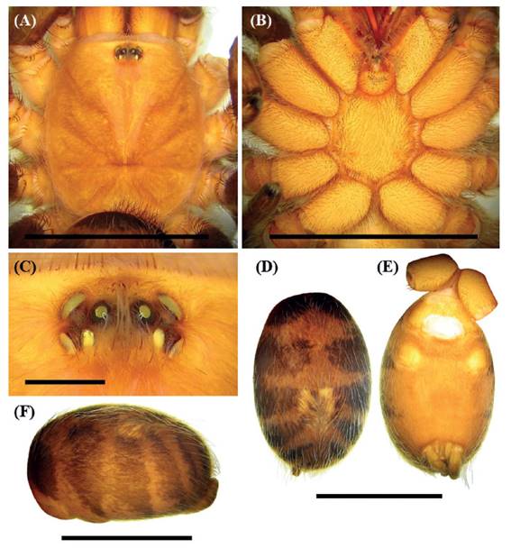
Figure 1 Anqasha picta (Pocock 1903), female (MUSM-ENT 0511132) from Peru, Ancash, Caraz. A. Carapace and coxae, dorsal view. B. Sternum, labium, maxillae and coxae, ventral view. C. Ocular tubercle, dorsal view. D-F. Abdomen with striped pattern. D. Dorsal view. E. Ventral view. F. Lateral view. Scale bar = 1 mm (Figure C). Scale bar = 10 mm (Figures A, B, D, E, F). Photo: Radan Kaderka.
Male palpal organ: short and stout embolus with PS, PI, A and R keel. PS keel developed and present on tegulum only, or absent, PI keel well-developed, bipartite, R and A keel weakly developed. Tegulum with distinct TP, projecting anteriorly (Sherwood & Gabriel 2022: figures 2 - 5; Figures 2, 6). Retrolateral lobe of cymbium weakly developed. Palpal tibia without retrolateral process, retrolateral face with or without a cluster of long spiniform setae. Two unequals tibial apophyses are present on tibia I (Sherwood & Gabriel 2022: figures 6 - 8; Figure 5B): a longer retrolateral tibial apophysis, usually with short apical spine and a shorter prolateral tibial apophysis usually with single, well-developed retrolateral spine at base. Metatarsus I slightly curved, without basal or median protuberance on retrolateral face. Male metatarsus I flexion on the apex of retrolateral branch.
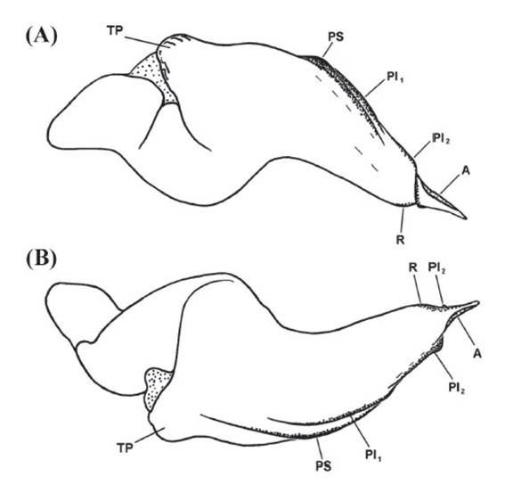
Figure 2 Anqasha picta (Pocock 1903), male (MUSM ENT 0511131) from Peru, Ancash, Yungay. Morphology of right palpal bulb. A. Prolateral view. B. Retrolateral view. PS = prolateral superior keel, PI1 = prolateral inferior keel, distal part, PI2 = prolateral inferior keel, proximal part, A = apical keel, R = retrolateral keel, TP = tegular protuberance (= TH, tegular heel sensu Sherwood & Gabriel (2022). Scale bar = 1 mm. Figured by Radan Kaderka.
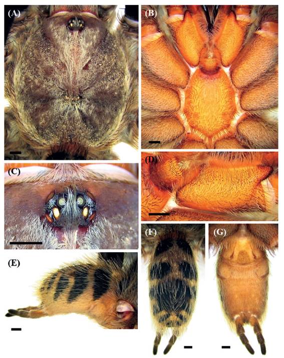
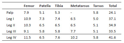
Figure 4 Anqasha minaperinensis sp. nov., male holotype (MUSM ENT 0509157) from Peru, Ancash, Huaraz, Mina Perina. A. Carapace, dorsal view. B. Sternum, labium, maxillae and coxae, ventral view. C. Ocular tubercle, dorsal view. D. Left maxilla and labium, ventral view. E-G. Abdomen with striped pattern. E. Lateral view. F. Dorsal view. G. Ventral view. Scale bar = 1 mm. Photo: Radan Kaderka.
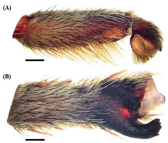
Figure 5 Anqasha minaperinensis sp. nov., male holotype (MUSM ENT 0509157) from Peru, Ancash, Huaraz, Mina Perina. A. Right palpal tibia, retrolateral view. B. Left tibia I, prolateroventral view. Scale bar = 1 mm. Photo: Radan Kaderka.
Females with spermathecae composed of two separated cylindrical seminal receptacles, distinctly granulated, with apical lobes and sclerotized basal plates, apical lobes are usually less sclerotized than basal plates (Sherwood & Gabriel 2022: figures 15 - 21; Figure 9).
Remarks. The following species were involved in the general description: male holotype of A. picta (BMNH), two females of A. minaperinensis sp. nov. (BMNH 1905.12.33 - 34), female of A. picta (MUSM-ENT 0511132), male holotype and female paratype of A. minaperinensis sp. nov. (MUSM-ENT 0509157).
Anqasha picta (Pocock, 1903)
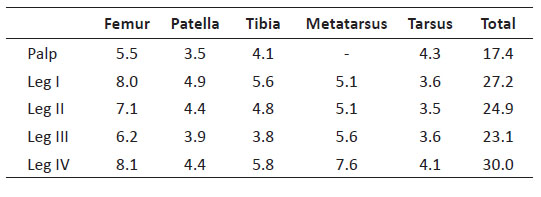
Table 1 Anqasha picta (Pocock 1903). Female (MUSM-ENT 0511132) from Caraz, Ancash, Peru. Lengths of palpal and leg segments (mm).
Hapalopus pictus: Pocock, 1903: 110. Male holotype, BMNH, from Caras, Peru, 2,200 m a.s.l., 12. December 1899, P. O. Simons col.
Homoeomma pictum: Gerschman de Pikelin & Schiapelli, 1973: 49, 54.
Anqasha picta: Sherwood & Gabriel, 2022: 248-253, figures 2-21.
Types. Male holotype (BMNH), from Peru, Ancash, Caras, 2,200 m a.s.l., 12 December 1899, The Andes, P. O. Simons col. Redescribed by Sherwood & Gabriel (2022): 248-249, figures 2-11.
Diagnosis. As per the generic diagnosis, Anqasha picta can be distinguished from other similar sympatric theraphosines based on genital organ morphology and the presence of prominent black banding on the dorsal and lateral opisthosoma (Sherwood & Gabriel, 2022).
Distribution and natural history. Known only from the valley of the Santa River in the province of Huaylas and Yungay in the region of Ancash, collected near Caraz and Yungay (Figure 10). The material from Yungay was obtained during daytime manual collecting in the zones between the grass and woodland up to 2600 m a.s.l., the temperature was of 20°C in the shade and about 14°C in ten centimeter long burrows under stones, with the relative humidity of 89% inside.
Remarks. Sherwood & Gabriel (2022) redescribed the male holotype including the palpal bulb morphology. After the careful examination of the published photos (Sherwood & Gabriel 2022: 2 - 5) I have revealed the erroneously named PS keel, which is in fact a short R keel, a keel developed retrolaterally from the apex of the embolus to the rear as defined by Bertani (2000). Prolateral keels (PS and PI keel) were defined by Bertani (2000) as two parallel keels, which are present in the prolateral area of the embolus (Bertani 2000). The homology of keels in the genus Anqasha is newly established (Figure 2).
The female from Caraz, the type locality of A. picta, is herein described for the first time. The morphology of seminal receptacles is different from those described by Sherwood & Gabriel (2022) in specimens collected near Recuay, Ancash, Peru.
The spermathecal morphology of specimens from Caraz and Yungay is also different in some aspects, e.g., the seminal receptacles in the female specimen from Yungay are cylindrical, more sclerotized and with less developed median constriction (Figures 9C, 9D). Both Caraz and Yungay are on the right bank of the Santa River, approximately 14 km distant from each other. Untill examples of female exuviae are collected and subjected to the comparative analysis, the specimens from Yungay (Figure 3) remain provisionally conspecific with A. picta.
FEMALE (MUSM-ENT 0511132) (Figures 1, 9A): Total length: 23.5, carapace length 10.0, width 8.5, chelicerae with 9 teeth on promargin, almost all the same size. Cheliceral denticulation from basal end: right side: V-V-VVVVVV-V, 9 big teeth. Left side: V-V-VVVVVV-V, 9 big teeth. Anterior eye row procurved, posterior eye row slightly recurved. Eye sizes and interdistances (Figure 1C): AME 0.34 (circular), ALE 0.44 (oval), PME 0.29 (oval), PLE 0.33 (oval), AME-AME 0.29, AME-ALE 0.13, PME-PME 0.70, PME-PLE 0.08, ALE-PLE 0.18, AME-PME 0.08, OQ length 0.88, width 1.56. Ocular tubercle low, almost undeveloped, length 1.17, width 1.56, clypeus narrow. Fovea transverse, slightly procurved, width 1.56, 6.9 from anterior edge of carapace. Labium length 1.35, width 1.40, anterior third with 43 cuspules, left and right maxilla with 158 and 163 cuspules. Sternum almost rounded, length 4.3, width 4.0, with three visible pairs of sternal sigilla located near coxae III (length 0.31, 0.44 from edge of sternum), coxae II (length 0.26, 0.18 from edge of sternum) and coxae I (length 0.16, 0.16 from edge of sternum). Leg formula: IV>I>II>III. All leg segments uniform. Maxillary and trochanteral stridulatory bristles absent.
Scopulae: All tarsi 100% densely scopulate, metatarsi I 100%, metatarsi II 85%, metatarsi III 30%, metatarsi IV 15% scopulate. Tarsal scopulae I-II integral, tarsal scopulae III divided by a band of setae, tarsal scopulae IV divided by a wide band of setae. Dorsal face of all tarsi and cymbium with two irregular longitudinal rows of very short claviform trichobothria. Denticulation of paired tarsal claws on right legs (prolateral / retrolateral row): I 5/4, II 4/4, III 6/5, IV 6/5. Plumose setae on retrolateral face of femur IV absent.
Spination: femora I p 0-0-1 (apical), II p 0-0-2 (apical), III 0, IV d 0-0-(0-1) (apical) and femora of palps 0; patellae I, II, III p 0-1-(0-1), IV 0 (apical) and patellae of palps 0; tibiae I v 0-1-0 (apical), II v (0-1)-1-(1-2) (apical), p 1-1-0 (apical), III v 2-(1-2)-(3-4) (apical), p 1-(1-3)-0 (apical), r 1-1-0 (apical), IV v (1-2)-(2-3)-3 (apical), p 0-0-1 (apical), r (0-1)-1-1 (apical) and tibiae of palps v (1-2)-1-3 (apical); metatarsi I v 1-(0-1)-2 (apical), II v (0-1)-(1-2)-3 (apical), p 0-1-1 (apical), III v (1-2)-(1-2)-(2-3) (apical), p (1-2)-2-2, r 1-1-1 (apical), IV v 1-2-3 (apical), p 0-(1-2)-(1-2) (apical), r 1-(2-3)-(1-2) (apical), tarsi I-IV and tarsi of palps 0.
Spermathecae (Figure 9A): two separated cylindrical seminal receptacles with sclerotized basal extension.
Abdomen: type III urticating setae are located in a dorsal rounded central patch. Length of patch: 3.56, width 3.56. Length of urticating setae 0.335-0.392. PLS: length 4.37, basal segment 2.03, middle segment 1.22, apical segment 1.12, all digitiform. PMS: 0.91.
Coloration and covering setae: dorsal view (Figure 1A): carapace covered with uniform yellowish-brown pubescence; coxae, trochantera and chelicerae with yellowish-brown pubescence; femora black with purple iridescence intermixed with long dark setae; patellae, tibiae, metatarsi and tarsi light brown intermixed with long pale setae. Prolateral face of coxae I without short spiniform setae. Abdomen (Figure 1D) covered with black setae intermixed with long pale setae, except for six pairs of yellowish-brown lateral stripes. All stripes dorsally converge, except for the third pair. The connection area of the first and second pair is anteriorly extended. The fourth, fifth and sixth pair are mutually joined on dorsal side. Abdominal long pale setae are more concentrated in longitudinal dorsal stripe than laterally. Ventral view (Figure 1B): labium, sternum, coxae and trochantera yellowish-brown, femora, patellae, tibiae and metatarsi light brown. Abdomen yellowish-brown without dark longitudinal band (Figure 1E). Spinnerets yellowish-brown.
Anqasha minaperinensis sp. Nov
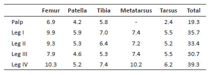
Table 2 Anqasha minaperinensis sp. nov. Male (MUSM-ENT 0509157) from Mina Perina, Ancash, Peru. Lengths of palpal and leg segments
Types. Male holotype, female paratype, immature female paratype (MUSM-ENT 0509157) from Peru, Department of Ancash, Huaraz, Mina Perina, 4,138 m a.s.l., December 15, 2015, A. Guzman col.
Etymology. A specific name refers to the name of the type locality near Mina Perina, Ancash, Peru.
Diagnosis. Males of Anqasha minaperinensis sp. nov. can be distinguished from A. picta by the shape of retrolateral branch of subapical apophyses on male tibia I (Figure 5B) which has a flattened apex having three short spines in A. minaperinensis sp. nov. (contra rounded apex with one subapical spine in A. picta). Females of A. minaperinensis sp. nov. differ from A. picta in the different shape of spermathecae (Figure 9). Both separate seminal receptacles have distinct granulation, long neck, apical extension, and sclerotized basal area. The apical lobes are less sclerotized than basal areas.
Distribution. Known only from the valley of the Santa River in the region of Ancash, collected near Mina Perina (correctly Mina Pierina) above Huaraz and near Recuay (Figure 10).
Remarks. Sherwood and Gabriel (2022) described two females (BMNH 1905.12.22-34; collected by P. O. Simons) coming from Paramo, Recuay (4000 m a.s.l.) in Peru as conspecific with Anqasha picta. As both females are significantly larger than the male holotype of A. picta, and they have different shape of spermathecae than the described female from the type locality in Caraz, both females (BMNH 1905.12.22-34) are herein transferred under A. minaperinensis sp. nov. based on the similarity of spermathecae and geographical distribution. The city of Recuay is approximatelly 87 km distant from the type locality of A. picta near Caraz and whatever gene flow between these populations is unlikely.
MALE (MUSM-ENT 0509157) (Figures 4-6): Total length: 23.82, carapace length 11.91, width 10.28, chelicerae with 11 teeth on promargin. Cheliceral denticulation from basal end: right side: v-V-v-V-VVVVVVv, 3 small and 8 big teeth. Left side: v-V-V-VVVVVV-VV, 1 small and 10 big teeth. Anterior eye row procurved, posterior eye row recurved. Eye sizes and interdistances (Figure 4C): AME 0.351 (circular), ALE 0.416 (oval), PME 0.299, PLE 0.299 (oval), AME-AME 0.312, AME-ALE 0.143, PME-PME 0.741, PME-PLE 0.130, ALE-PLE 0.364, AME-PME 0.078, OQ length 0.923, width 1.560. Ocular tubercle length 1.378, width 1.560, clypeus narrow. Fovea transverse, deep, procurved, width 1.58, 7.70 from anterior edge of carapace. Labium length 1.575, width 1.981, anterior third with 39 cuspules, left and right maxilla with 178 and 204 cuspules in basal half, apical half with short spiniform setae. Maxillary lobe well-developed, elongated (Figure 4D). Labiosternal sigilla joined. Sternum length 6.224, width 4.699, three pairs of sternal sigilla located near coxae III (length 0.468, 0.702 from edge of sternum), coxae II (length 0.364, 0.234 from edge of sternum) and coxae I (length 0.247, 0.247 from edge of sternum) (Figure 4B). Leg formula: IV>I>II>III. Incrassate leg segments: slightly incrassate femur III. Maxillae and trochantera without stridulatory bristles.
Scopulae: All tarsi 100% densely scopulate, metatarsi I 100%, metatarsi II 60%, metatarsi III 30%, metatarsi IV 25% scopulate. Tarsal scopulae I undivided, tarsal scopulae II divided by a longitudinal line of setae, tarsal scopulae III-IV divided by a longitudinal band of setae. Dorsal face of all tarsi and cymbium with two irregular longitudinal rows of very short claviform trichobothria. Denticulation of paired tarsal claws on right legs (prolateral / retrolateral row): I 3/2, II 3/5, III 3/3, IV 5/- (missing claw). Plumose setae on retrolateral face of femur IV absent.
Spination: femora I d 0-1-2, II p 0-2-0, d 0-0-1, III p 0-2-1, d 0-0-1, IV p 0-2-1, d 0-0-1 and femora of palps p 0-0-1; patellae I v 0-0-2 (apical), II v 0-0-2 (apical), p 0-1-1, III v 0-0-1 (apical), p 0-1-1, IV p 0-3-0 and patellae of palps 0; tibiae I v 6-5-2 (apical), p 1-0-1, r 2-1-1, II v 4-4-7 (5 apical), p 0-1-1, r 3-2-2, III v 3-4-4 (apical), p 2-1-0, r 2-1-1, d 3-0-0, IV v 5-3-3 (apical), p 2-1-1, r 1-2-1, d 2-0-0 and tibiae of palps p 1-2-1; metatarsi I v 0-2-3 (apical), p 1-1-1, r 2-1-1, II v 3-4-2 (apical), p 1-1-1, r 3-2-2, III v 3-2-2-6 (apical), p 1-1-0, d 2-2-4, IV v 5-3-3-4 (apical), p 1-2-2, r 2-1-1, d 0-1-2, tarsi I-IV and tarsi of palps 0.
Palpal organ as in Figure 6, embolus short and stout with four keels, PS, PI, A and R keel, PI keel is bipartite, its distal part on embolus is sigmoidly curved and without semi-circular or subtriangular lobe. PS keel, restricted to tegulum only, short and parallel with the proximal part of PI keel. A keel transparent and developed. R keel short, located near apex and parallel with PI keel. Tegulum with subconical apophysis, projecting anteriorly. Retrolateral lobe of cymbium, which is smaller than prolateral lobe, covered with short spiniform setae. Palpal tibia distally tapering. Retrolateral face of palpal tibia without subapical process but with a group of long spiniform setae (Figure 5A). Two unequal subapical apophyses are present on tibia I (Figure 5B): a longer retrolateral tibial apophysis with flattened apex having three short apical spines, a shorter prolateral tibial apophysis with single, retrolateral spine reaching the apex, approximately of two third of the length of prolateral tibial apophysis. Metatarsus I slightly curved and without basal or median protuberance on retrolateral face. When flexed, metatarsus I contacts the apex of retrolateral tibial apophysis.
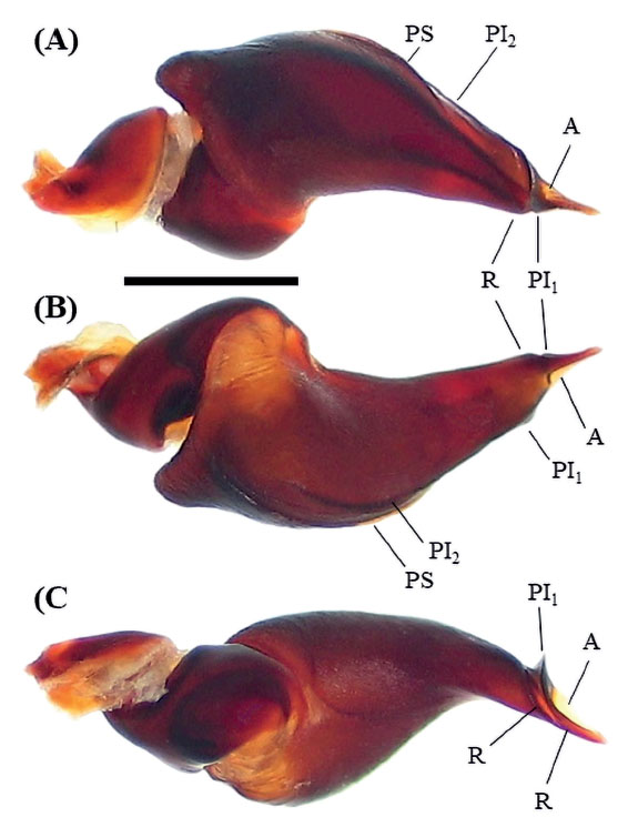
Figure 6 Anqasha minaperinensis sp. nov., male holotype (MUSM ENT 0509157) from Peru, Ancash, Huaraz, Mina Perina. Morphology of right palpal bulb. A. Prolateral view. B. Retrolateral view. C. Ventral view. PS = prolateral superior keel, PI1 = prolateral inferior keel, distal part, PI2 = prolateral inferior keel, proximal part, A = apical keel, R = retrolateral keel, TP = tegular protuberance. Scale bar = 1 mm. Photo: Radan Kaderka.
Abdomen (Figure 4E-G): type III urticating setae are located in almost circular dorsocentral patch. Size of the patch: length 4.43, width 4.17. PLS: length 4.82, basal segment 2.11, middle segment 1.25, apical segment 1.46, all digitiform. PMS: 1.25.
Coloration and covering setae: dorsal view (Figures 4A, 4F): carapace and chelicerae covered with yellowish pubescence; coxae, trochantera yellowish; femora black; patellae, tibiae, metatarsi and tarsi brown. Patellae I and II with two equal longitudinal glabrous stripes, patellae III, IV with one such diagonal glabrous stripe. Abdomen black with five lateral yellowish stripes, except for the dorsocentral dark brown patch of type III urticating setae. Ventral view: brown, except for ventral abdomen which is yellowish (Figure 4G). Spinnerets: PLS: apical and middle segment dark brown, basal segment distally dark brown, proximally light brown. AMS: basal two thirds light brown, apical third dark brown.
FEMALE (MUSM-ENT 0509157) (Figure 7): Total length: 37.23, carapace length 16.53, width 13.96, chelicerae with 9-10 teeth on promargin. Cheliceral denticulation from basal end: right side: V-V-VVVVVVVV, 10 big teeth. Left side: V-V-VVVVVVV, 9 big teeth. Anterior eye row slightly procurved, posterior eye row recurved. Eye sizes and interdistances (Figure 7C): AME 0.39 (circular), ALE 0.49 (oval), PME 0.38 (oval), PLE 0.40 (oval), AME-AME 0.35, AME-ALE 0.22, PME-PME 0.91, PME-PLE 0.08, ALE-PLE 0.27, AME-PME 0.17, OQ length 1.053, width 2.002. Ocular tubercle: length 1.768, width 2.002, clypeus narrow. Fovea transverse, deep, slightly procurved, width 2.44, 11.39 from anterior edge of carapace. Labium length 2.19, width 2.85, anterior third with 43 cuspules, left and right maxilla with 233 and 262 cuspules in basal half and short spiniform setae in apical half (Figure 7D). Sternum length 7.74, width 6.58, with three visible pairs of sternal sigilla located near coxae III (length 0.52, 1.01 from edge of sternum), coxae II (length 0.40, 0.60 from edge of sternum) and coxae I (length 0.23, 0.61 from edge of sternum). Leg formula: IV>I>II>III. All leg segments uniform.
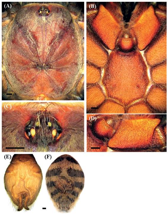
Figure 7 Anqasha minaperinensis sp. nov., female paratype (MUSM ENT 0509157) from Peru, Ancash, Huaraz, Mina Perina. A. Carapace, dorsal view. B. Sternum, labium, maxillae and coxae, ventral view. C. Ocular tubercle, dorsal view. D. Left maxilla and labium, ventral view. E-F. Abdomen with striped pattern. E. Ventral view. F. Dorsal view. Scale bar = 1 mm. Photo: Radan Kaderka.
Table 3 Anqasha minaperinensis sp. nov. Female (MUSM-ENT 0509157) from Mina Perina, Ancash, Peru. Lengths of palpal and leg segments
Scopulae: All tarsi 100% densely scopulate, metatarsi I 100%, metatarsi II 65%, metatarsi III 40%, metatarsi IV 25% scopulate. Tarsal scopulae I, II integral, tarsal scopulae III, IV divided by a longitudinal band of setae. Dorsal face of all tarsi and cymbium with two irregular longitudinal rows of very short claviform trichobothria. Denticulation of paired tarsal claws on left legs (prolateral / retrolateral row): I 2/missing, II 4/2, III 5/5, IV 5/5, undeveloped. Plumose setae on retrolateral face of femur IV absent.
Spination: femora I, II and III p 0-0-1 and femora of palps p 0-0-1, patellae I-IV and patellae of palps 0, tibiae I p 0-1-0, II v 0-1-2 (apical), p 0-1-0, III v 1-2-3 (apical), p 1-1-0, r 1-1-0, IV v 2-2-3 (apical), r 1-1-1 and tibiae of palps v 1-2-2 (apical), p 0-1-0, metatarsi I 0, II v 1-1-2 (apical), III v 3-2-5 (apical), p 0-1-1, r 0-1-1, IV v 3-2-3 (apical), p 0-1-1, r 1-1-2, tarsi I-IV and tarsi of palps 0.
Spermathecae (Figure 9E, 9F): two separated seminal receptacles, each with distinct granulation, long tubular neck, apical extension and sclerotized basal area, apical lobes less sclerotized than basal areas.
Abdomen: type III urticating setae are located in dorsocentral oval patch. Size of the patch: length 4.4, width 5.5. PLS: length 5.59, basal segment 2.39, middle segment 1.20, apical segment 2.00, all digitiform. PMS: 1.64.
Coloration and covering setae: dorsal view (Figure 7A): carapace dark brown with short pale covering setae; coxae and trochantera light brown; chelicerae light brown; femora black; patellae, tibiae, metatarsi and tarsi brown, intermixed with long pale setae. Patellae I, II and palpal patella with two almost equal parallel longitudinal glabrous stripes, patellae III, IV with two unequal diagonal stripes. Palpal femur prolaterally bare. Femora I-IV retrolaterally with a narrow longitudinal glabrous stripe. Abdomen (Figure 7F) light brown with five well-developed black dorsolateral stripes, one pair of stripes near spinnerets are less developed. Ventral view (Figure 7B): labium and maxillae reddish-brown; sternum, coxae, trochantera brown; femora dark brown; patellae, tibiae and metatarsi brown. Abdomen ventrally yellowish brown without dark longitudinal band (Figure 7E). Spinnerets yellowish brown.
Variability. The variability in morphology of spermathecae in Figure 9.
Anqasha sp.
Distribution. Known only from Lomas Flor de Amancaes, the mountain ridge in the central part of Lima reaching the altitude of over 700 m a.s.l. (Figure 10).
FEMALE (MUSM-ENT 0501413, probably immature) (Figure 8): Total length: 13.31, carapace length 5.65, width 4.34, chelicerae with 9 teeth on promargin. Cheliceral denticulation from basal end: right side: V-V-VVVVVV-v, 1 small and 8 big teeth. Left side: V-V-vvvVV-V-v, 4 small and 5 big teeth. Anterior eye row slightly procurved, posterior eye row recurved. Eye sizes and interdistances (Figure 8C): AME 0.23 (circular), ALE 0.32 (oval), PME 0.18 (oval), PLE 0.24 (oval), AME-AME 0.04, AME-ALE 0.09, PME-PME 0.44, PME-PLE 0.03, ALE-PLE 0.10, AME-PME 0.03, OQ length 0.57, width 0.96. Ocular tubercle: length 0.75, width 0.96, clypeus narrow. Fovea transverse, procurved, width 0.83, 4.00 from anterior edge of carapace. Labium length 0.70, width 1.17, anterior third with 14 cuspules, maxillae with 79-80 cuspules in basal half and short sparse spiniform setae in apical half. Sternum length 2.79, width 2.38, with three visible pairs of sternal sigilla located near coxae III (length 0.10, 0.27 from edge of sternum), coxae II and coxae I. Leg formula: IV>I>II>III. All leg segments uniform.
Figure 8 Anqasha sp., female (MUSM-ENT 0501413) from Peru, Lima, Lomas Flor de Amancaes, 470-732 m a.s.l. A. Carapace and coxae, dorsal view. B. Sternum, labium, maxillae, coxae and trochantera, ventral view. C. Ocular tubercle, dorsal view. D. Left maxilla and labium, ventral view. E-F. Abdomen with striped pattern. E. Dorsal view. F. Ventral view. Scale bar = 1 mm. Photo: Radan Kaderka.
Table 4 Anqasha sp. Female (MUSM-ENT 0501413) from Lomas Flor de Amancaes, Lima, Peru. Lengths of palpal and leg segments:

Scopulae: All tarsi 100% densely scopulate, metatarsi I 50%, metatarsi II 30%, metatarsi III, IV ascopulate. Tarsal scopulae I, II divided by a longitudinal band of setae, in tarsi III, IV divided by a wide band of setae. Dorsal face of all tarsi and cymbium with two irregular longitudinal rows of very short claviform trichobothria. Denticulation of paired tarsal claws on right legs (prolateral / retrolateral row): I 3/3, II 3/3, III 3/3, IV 4/3. Plumose setae on retrolateral face of femur IV absent.
Spination: femora I, II and III d 0-0-1 and femora of palps d 0-0-1, patellae I-IV and patellae of palps 0, tibiae I 0, II v 0-1-0, p 0-1-0, III v 0-2-2 (apical), p 1-1-0, r 1-1-0, IV v 1-2-2 (apical), r 0-1-1 and tibiae of palps v 0-1-2 (apical), metatarsi I v 0-1-2 (apical), II v 0-1-3 (apical), III v 0-2-3 (apical), p 1-1-1, r 0-1-1 (apical), IV v 2-1-3 (apical), p 0-1-1 (apical), r 1-2-1, tarsi I-IV and tarsi of palps 0.
Spermathecae (Figure 9B): two separated seminal receptacles with distinct apical granulation and sclerotized basal extension.
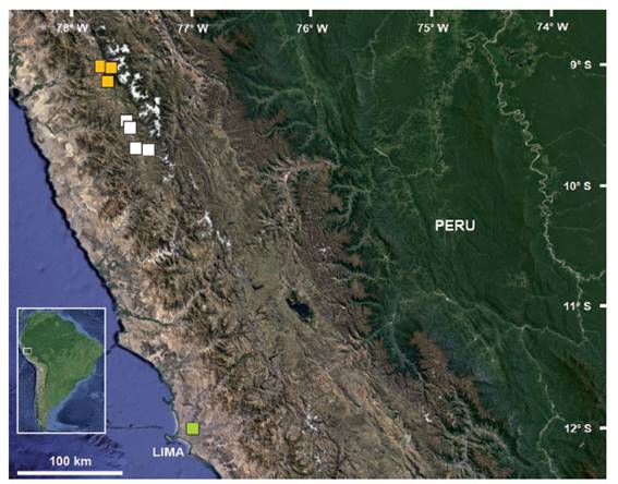
Figure 9 A. Anqasha picta (Pocock 1903), female (MUSM-ENT 0511132) from Peru, Ancash, Caraz, morphology of spermathecae, dissected from specimen in alcohol. B. Anqasha sp., female (MUSM-ENT 0501413) from Peru, Lima, Lomas Flor de Amancaes, morphology of spermathecae, dissected from specimen in alcohol. C. Anqasha picta (Pocock 1903), female (MUSM-ENT 0511130) from Peru, Ancash, Yungay, morphology of spermathecae, dissected from exuvia. D. Anqasha picta (Pocock 1903), female (MUSM-ENT 0511130) from Peru, Ancash, Yungay, morphology of spermathecae, dissected from specimen in alcohol. E. Anqasha minaperinensis sp. nov., female paratype (MUSM-ENT 0509157, bigger specimen) from Peru, Ancash, Mina Perina, morphology of spermathecae, ventral view, dissected from specimen in alcohol. F. Anqasha minaperinensis sp. nov., female paratype (MUSM-ENT 0509157, smaller specimen) from Peru, Ancash, Mina Perina, morphology of spermathecae, ventral view, dissected from specimen in alcohol. Scale bar = 1 mm. Photo: Radan Kaderka.
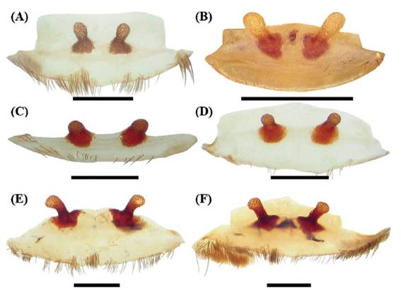
Figure 10 Distribution map of the genus Anqasha in Peru. Yellow rectangle for Anqasha picta. White rectangle for Anqasha minaperinensis sp. nov. Green rectangle for Anqasha sp. Prepared by Radan Kaderka.
Abdomen: type III urticating setae are located in small oval dorsocentral patch. PLS: length 2.47, basal segment 1.12, middle segment 0.60, apical segment 0.75, all digitiform. PMS: 0.65.
Coloration and covering setae: dorsal view (Figure 8A): carapace brown, coxae and trochantera brown; chelicerae brown; femora dark brown; patellae, tibiae, metatarsi and tarsi brown, intermixed with long pale setae. Abdomen (Figure 8E) black, intermixed with long pale setae, and dorsally and laterally with 4-5 transverse yellowish stripes. Length of central patch: 0.52, width 1.12. Ventral view (Figure 8B): labium, sternum, coxae, maxillae, trochantera, femora, patellae, tibiae and metatarsi yellowish brown. Abdomen ventrally without dark longitudinal band (Figure 8F). Spinnerets yellowish brown.

