SciELO - Scientific Electronic Library Online

 
vol.39 número4Linfangiomatosis esplénica en un paciente adulto: Reporte de casoTerminología en educación médica a distancia índice de autoresíndice de materiabúsqueda de artículos
Home Pagelista alfabética de revistas  

Servicios Personalizados

Revista

Articulo

Indicadores

  • No hay articulos citadosCitado por SciELO

Links relacionados

  • No hay articulos similaresSimilares en SciELO

Compartir


Acta Médica Peruana

versión On-line ISSN 1728-5917

Acta méd. Peru vol.39 no.4 Lima oct./dic. 2022

http://dx.doi.org/10.35663/amp.2022.394.2334 

Reporte de casos

Patient with Rosai-Dorfman-Destombes disease:18F-FDG-PET/CT as a diagnostic tool

Paciente con enfermedad de Rosai-Dorfman-Destombes: 18F-FDG-PET/CT como herramienta diagnóstica

Luis C. Araujo Cachay1  , Médico-cirujano, Especialista en Medicina Nuclear, Doctor en Medicina
http://orcid.org/0000-0002-3609-0517

1Departamento de Medicina Nuclear, Instituto Nacional de Enfermedades Neoplásicas (INEN), Lima-Perú

2 Facultad de Medicina, Universidad Ricardo Palma, Lima-Perú

ABSTRACT

Rosai-Dorfman-Destombes disease (RDD) is a rare non-Langerhans cell histiocytosis, with sinus involvement and massive lymphadenopathy. RDD is usually self-limited; it can appear alone or related to other diseases. We present a 9-year-old male with biopsy of that lesion was taken, giving as result a benign histiocytosis, compatible with RDD. The patient was scanned with full body 18F-FDG PET/CT, the results of the study showed hypermetabolic focal lesions in the sphenoid sinus, ethmoid sinus, and bilateral nasal involvement with hypermetabolic focal injury in the middle third of the left tibia, findings in relation to high-grade expansive injury. The study of 18F-FDG PET/CT demonstrated avid FDG lesions at nodal and extranodal sites, and it also can be used in monitoring and/or evaluating response to treatment.

RESUMEN

La enfermedad de Rosai-Dorfman-Destombes (RDD) es una rara histiocitosis de células no Langerhans, con compromiso de los senos nasales y linfadenopatías masivas. RDD es usualmente autolimitada, y puede aparecer sola o asociada a otras enfermedades. Se presenta el caso de un niño varón de 9 años cuyo resultado histopatológico mostró histiocitosis benigna compatible con RDD. El paciente se sometió a una Tomografía Computarizada/Tomografía de Emisión de Positrones con 18F-fluorodeoxyglucosa (18F-FDG-TEP/TC) de todo el cuerpo, que mostró lesiones focales hipermetabólicas en los senos esfenoidales, etmoidales, y nasales bilaterales, junto a una lesión focal hipermetabólica en el tercio medio de la tibia izquierda. El estudio con 18F-FDG-PET/CT pudo demostrar lesiones ávidas de glucosa en sitios nodales y extra-nodales, y también puede servir en la monitorización o evaluación de la respuesta al tratamiento.

INTRODUCTION

Rosai-Dorfman-Destombes disease (RDD) is a rare non-Langerhans cell histiocytosis, with sinus involvement and massive lymphadenopathy 1). RDD prevalence is 1:200 000, having a predilection for men, the mean age of presentation is 20,6 years, being more frequent in children 2). RDD is usually self-limited; it can appear alone or related to other diseases (autoimmune or malignancy) 3. Extranodal disease can present in 43 % patients 3). The involvement of head/neck is described in 11 % of patients producing symptoms due to mass effect 4,5). Bone involvement occurs in 5 % to 10 %, the most common symptom is bone pain, however pathologic fracture can occur (diaphysis and metaphysis affection) 4). The published cases of 18F-FDG PET/CT in the RDD, report avidity of FDG radiotracer by the injuries, this is a powerful diagnostic tool; a full body scan can diagnose with high specificity the extranodal involvement 6). The purpose of this clinical case is to describe the metabolic characteristics of 18F-FDG-PET/CT in Rosai-Dorfman-Destombes disease.

CASE REPORT

We present a 9-year-old male, with 5-months characterized by headache in the left side of the head, eye pain, obstruction of the left nostril, explosive vomiting, mouth breathing, and nighttime snoring. In the physical exam, the main finding was a mass in the left nostril occluding the entire lumen. A biopsy of that lesion was taken, giving as result a benign histiocytosis, compatible with RDD. Subsequently the patient was scanned with full body 18F-FDG PET/CT, the image acquisition after 60 minutes, PET/CT equipment (Philips Gemini TF). The results of the study showed hypermetabolic focal lesions in the sphenoid sinus, ethmoid sinus, and bilateral nasal involvement, SUVmax in 8.3, increasing to 13.8 in late control, associated with hypermetabolic focal injury in the middle third of the left tibia with SUVmax in 9.6, findings in relation to high-grade expansive injury (Figure 1). Multiple bilateral cervical lymphadenopathies with SUVmax in 7.0 suggestive of nodal involvement. The patient was started with a combined treatment consisting of 6-MP, methotrexate, and vinblastine.

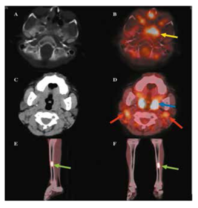
Figure 1.  18F-FDG-PET/CT Results: Axial CT (A) and axial fused (B) images showing focal hypermetabolic intake in the sphenoid lesion (yellow arrow). Axial CT (C) and axial fused (D) images showing focal hypermetabolic intake in the bilateral nasal cavity (blue arrow) and bilateral latero-cervical nodes (red arrow). Sagittal CT fused (E) and coronal CT fused (F) images showing focal hypermetabolic intake in the medial diaphysis of the left tibia (green arrows). 

DISCUSSION

RDD has been described a few thousand times in all medical records. The typical clinical presentation of this patient was helpful for establishing the diagnosis. Nevertheless, RDD can be confused with many other diseases that produce generalized lymphadenopathy, from infectious etiologies to neoplasm. A misleading diagnosis would cause harmful outcomes due to inadequate treatment. In the setting of a low-income country as Peru, this case commonly can be confused with Mycobacterium tuberculosis infection, delaying the RDD treatment. In early 2018, there was an effort to create diagnostic criteria and treatment guidelines to unify the disease management. In Abla O, et al. 2018, mention that some authors recommend PET/CT scan for all patients, the study of 18F-FDG PET/CT as part of the diagnostic process demonstrated avid FDG lesions at nodal and extranodal sites. It is evident that 18F-FDG PET/CT not only locates the disease but can be used in monitoring and/or evaluating response to treatment. More studies are required to determine the exact role of 18F-FDG PET/CT in this pathology.

Bibliography

1. Rosai J, Dorfman RF. Sinus histiocytosis with massive lymphadenopathy: a newly recognized benign clinicopathological entity. Arch Pathol. 1969;87(1):63-70. [ Links ]

2. Mahzoni P, Zavareh MH, Bagheri M, Hani N, Moqtader B. Intracranial ROSAI-DORFMAN disease. J Res Med Sci. 2012;17(3):304-307. PMID: 23267385; PMCID: PMC3527051. [ Links ]

3. Abla O, Jacobsen E, Picarsic J, Krenova Z, Jaffe R, Emile J-F, et al. Consensus recommendations for the diagnosis and clinical management of Rosai-Dorfman-Destombes disease. Blood. [Internet]. Junio del 2018 [Citado el 01 de agosto del 2019];131(26):2877-90. DOI: 10.1182/blood-2018-03-839753 [ Links ]

4. Foucar E, Rosai J, Dorfman R. Sinus histio- cytosis with massive lymphadenopathy (Rosai-Dorfman disease): review of the entity. Semin Diagn Pathol. 1990;7(1):19-73. PMID: 2180012. [ Links ]

5. Vujhini SK, Kolte SS, Satarkar RN, Srikanth S. Fine needle aspiration diagnosis of Rosai-Dorfman Disease involving thyroid. J Cytol. 2012 Jan;29(1):83-5. doi: 10.4103/0970-9371.93239. [ Links ]

6. Albano D, Bosio G, Bertagna F. 18F-FDG PET/CT Follow-up of Rosai-Dorfman Disease. Clin Nucl Med. 2015 Aug;40(8):e420-2. doi: 10.1097/RLU.0000000000000853. [ Links ]

Contribution Details:LCAC conceived the research idea, gathered the clinical information and images, wrote and edit the manuscript, and approved the final version submitted to the journal.

Funding:The author did not receive any funding for this research.

Citar como: Paciente con enfermedad de Rosai-Dorfman- Destombes: 18F-FDG-PET/TC como herramienta diagnóstica. Acta Med Peru. 2022; 39(4): 396-8. doi: https://doi.org/10.35663/amp.2022.394.2334

Received: February 23, 2022; Accepted: October 06, 2022

Correspondencia Luis C. Araujo Cachay E-mail: laraujo@inen.sld.pe

Conflict of interest: The author does not have any conflicts of interest.

Creative Commons License This is an open-access article distributed under the terms of the Creative Commons Attribution License