SciELO - Scientific Electronic Library Online

 
vol.40 issue4Randomized Controlled Clinical Trial of Two Surgical Techniques for Primary Pterygium in Jungle Patients: Cauterization versus SuturingUnusual case of diffuse B lymphoma of large cells (NOS) primary cerebral associated with hydrocephaly and neurosurgical management author indexsubject indexarticles search
Home Pagealphabetic serial listing  

Services on Demand

Journal

Article

Indicators

  • Have no cited articlesCited by SciELO

Related links

  • Have no similar articlesSimilars in SciELO

Share


Acta Médica Peruana

On-line version ISSN 1728-5917

Acta méd. Peru vol.40 no.4 Lima Oct./Dec. 2023  Epub Dec 31, 2023

http://dx.doi.org/10.35663/amp.2023.404.2662 

Reporte de casos

Candida parapsilosis endocarditis without vegetations at Echocardiography: a case report

Endocarditis por Candida parapsilosis sin vegetaciones ecocardiográficas: Un reporte de caso

Natalia Vargas-Herrera1  2  *  , Médico infectólogo
http://orcid.org/0000-0002-0948-0114

Jose M. Estrada-Grossmann3  , Médico cirujano
http://orcid.org/0000-00028618-3359

Marcos Saavedra-Velasco4  , Médico infectólogo
http://orcid.org/0000-0003-2977-1020

Gian Marco Arango-Rojas5  , Médico infectólogo
http://orcid.org/0000-0002-8396-6227

John Cabrera-Enríquez6  7  , Médico infectólogo
http://orcid.org/0000-0002-0770-505X

1 Centro Nacional de Salud Pública. Unidad de intervenciones estratégicas.

2 Instituto Nacional de Salud, Lima Perú

3 Universidad Peruana de Ciencias Aplicadas, Lima Perú.

4 Instituto de Investigación en Ciencias Biomédicas (INICIB). Universidad Ricardo Palma, Lima-Perú

5 Servicio de infectología, Hospital Nacional Edgardo Rebagliati Martíns Lima-Perú

6 Carrera de Medicina Humana, Ciencias de la Salud. Universidad San Ignacio de Loyola, Lima-Perú

7 Servicio de Clínica Pediátrica, Hospital Nacional Guillermo Almenara Yrigoyen. Lima-Perú

ABSTRACT

Candida endocarditis is a very serious manifestation of candida infections, and it has increased in incidence over the past years. Of these, C. parapsilosis has been described as a cause of endocarditis in native valves of intravenous drug users and prosthetic valves. We report the case of a female that developed a cerebrovascular accident secondary to emboli from aortic prosthetic valve C. parapsilosis endocarditis, despite apparently normal echocardiography. She received antifungal therapy without surgical intervention.

Key words: Candida parapsilosis; Endocarditis; Echocardiography, Transesophageal; Biofilms; Candidiasis, Invasive

RESUMEN

La endocarditis por Candida spp. es una manifestación muy grave de las infecciones por este patógeno y su incidencia ha ido aumentando en los últimos años. La Candida parapsilosis se ha descrito como causa de endocarditis en válvulas nativas de usuarios de drogas intravenosas y prótesis valvulares. Presentamos el caso de una mujer que desarrolló un accidente cerebrovascular secundario a émbolos fúngicos procedentes de una prótesis valvular aórtica infectada por C. parapsilosis, a pesar de un ecocardiograma sin vegetaciones. La paciente recibió tratamiento antifúngico sin necesidad de intervención quirúrgica.

Palabras clave: Candida parapsilosis; Endocarditis; Ecocardiografía Transesofágica; Biopelículas; Candidiasis Invasiva

INTRODUCTION

C. parapsilosis has been considered a nosocomial pathogen for years, it frequently colonizes skin (it has been isolated in the hands of healthcare professionals) and adheres to synthetic material such as parenteral nutrition lines, intravascular catheters and other prosthetic devices by using biofilms1,2.

Among fungal endocarditis, C. parapsilosis represents the most common non-albican species isolated, and it is known to be an important cause of invasive candidiasis2,3. Heart valve surgery, intravenous drug consumption, prolonged antibiotic therapy, immunosuppression, previous episodes of endocarditis, and preexisting valve disease, are some of the main predisposing factors for C. parapsilosis infective endocarditis(CPIE)3,4. Of note, the most commonly affected heart tissue is the aortic valve, followed by the mitral and the tricuspid valve; also, in the outbreaks of CPIE, it has been found that intraoperative contamination is the most possible source of infection4.

In the literature there are some case reports of CPIE but most of these have imaging evidence of vegetations on the echocardiography. We describe the case of a middle-aged peruvian woman with history of heart valve surgery who presented to the emergency with a cardioembolic event due to a fungal endocarditis despite four echocardiographic procedures without evidence of vegetations or valve lesions. Treatment with high dose echinocandin was successful without the need of surgical intervention.

CASE REPORT

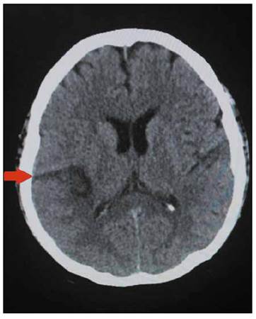
A 49-year-old female with past medical history of hypertension and severe aortic stenosis with subsequent aortic valve placement by modified Bentall procedure 8 months before, presented to the hospital with a seven-hour history of left arm and left leg numbness plus weakness and dysarthria. She was using warfarin since her prosthetic valve surgery. On admission, she complained of occipital headache and showed left hemiplegia on physical examination; no mental status changes or heart murmurs were auscultated. She was afebrile with a temperature of 36.5°C, a heart rate of 98 beats per minute, blood pressure of 140/90mmHg and respiratory rate of 18 breaths per minute. The brain computed tomography (CT) showed an ischemic stroke in the territory of the right middle cerebral artery (Figure 1), however no atherosclerosis was found on the carotid doppler ultrasound, and the trans-thoracic echocardiography (TTE) did not show vegetations. Additionally, the electrocardiogram was unremarkable.

Figure 1 Non-contrast brain computed tomography showing an ischemic lesion (hypodense area) 

Her white blood cell count was 5.48 cells/mm3, hemoglobin of 11.9 g/dL and she had a normal platelet count of 201 x 109/L. She was subsequently admitted to the neurology ward.

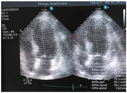
On the third day of hospitalization, she began with daily temperatures of 37.6°C that reached 38.9°C plus episodes of hypotension, she was started on empiric intravenous (IV) Vancomycin 1g/12h. plus Ceftriaxone 2g/day. Her urine analysis and chest radiograph where unremarkable, however she was still febrile, and her third set of blood cultures started growing yeast; the patient was started on IV Fluconazole 200mg/12h. Since a second TTE was normal, a transesophageal echocardiography (TEE) (Figure 2) was performed on the second week of hospitalization given the high suspicion of endocarditis but did not report any cardiac lesions including fistula or dehiscence. Blood cultures pending results confirmed the presence of C. parapsilosis in addition, a CT abdomen and eye examination were unremarkable.

Figure 2 Transesophageal echocardiography not showing vegetations 

During a 2-week period many infectious agents were tested including HBV, HCV, HIV, HTLV, tuberculosis and there were no markers of autoimmune disease. Despite antifungal therapy with IV fluconazole for almost 2 weeks, in which isolated C. parapsilosis was susceptible to all antifungals tested, except to ketoconazole according to the antifungal sensitivity test, MicroScan® (according to the FDA, is an automated system that provides results that are substantially equivalent to the generated using suitable reference methods)5 , the patient remained febrile, presented again episodes of hypotension and her INR reached 5.7. On the third week, in view of a possible interaction between warfarin and fluconazole, this antifungal was switched to Caspofungin 70mg IV as first day loading dose, followed by 50mg/day IV. During that week, the patient did not present fever again, was hemodynamically stable but her new set of blood cultures yielded C. parapsilosis once again on the tenth day of Caspofungin therapy. A second TEE was performed searching for fungal vegetations that could explain the persistent fungemia despite the prolonged antifungal therapy, but once again it was unremarkable. Caspofungin dose was increased to 150mg/day IV (invasive candidiasis dose according to the 2016 IDSA guidelines) for 1 week. Control blood cultures repeated daily from day 1 of the high Caspofungin dose were sterile and patient did not develop fever again.

She was finally discharged on oral Fluconazole 14 days after her last sterile blood culture, asymptomatic and hemodynamically stable. The patient was scheduled for follow-up in the infectious diseases clinic

DISCUSSION

The frequency of endocarditis due to C. parapsilosis has increased in recent years, this due to its ability to form biofilms which also explains its high mortality1, and according to the 2023 Duke ISCVIV Criteria, Candida spp. is now considered a "typical" infective endocarditis (IE) pathogen in the setting of intracardiac prosthetic material6.

For a successful outcome, an accurate diagnosis of IE is necessary to provide adequate treatment. In this case, the patient had two positive C. parapsilosis blood cultures which constitute a major criterion plus three minor criteria for IE: previous history of prosthetic valve, previous valve repair and fever. This confirmed the diagnosis.

On the other hand, despite having several echocardiographic procedures including TEE (which has high specificity and sensitivity especially in patients with prosthetic valves)7, no vegetations were identified. This could be explained by the fact that fungal vegetations have a higher incidence of embolic events compared to bacterial endocarditis, and these may had detached before TEE was performed8. In addition, it has been described that TEE could not confirm or exclude an IE diagnosis in challenging clinical situations such as patients with intracardiac implants6. In those scenarios, other diagnostic techniques such as cardiac computed tomography (CCT) and/or positron emission computed tomography (PET/CT) should be used in order to improve the identification of IE especially of prosthetic origin 6. In fact, CCT in combination with TEE has higher sensitivity for the detection of both valvular and paravalvular lesions, while PET/CT is preferred to diagnose PVE over echocardiography. Both imaging techniques are included in the 2023 Duke-ISCVID IE Criteria9.

Regarding clinical manifestations, common infectious endocarditis signs like heart murmurs may be absent in fungal endocarditis, however Candida endocarditis has certain characteristics such as loss of visual acuity or cutaneous nodules. Likewise, the vegetations are large and have a greater tendency to embolize7. Also, Garzoni in his review of the literature regarding C. parapsilosis endocarditis, found that 43.8% of 72 cases presented cerebral thromboembolic events, and he also mentioned that fever and embolic events should raise the suspicion of infective endocarditis, specially of fungal origin9. In addition, Candida spp is described in the literature as a cause of cardioembolic strokes and Lipton et al mentioned in their study that when Candida endocarditis is suspected, the rate of spread to the central nervous system can reach up to 80%10.

The definitive treatment for Candida endocarditis is the combination of antifungal therapy and surgery according to the 2016 IDSA candidiasis guidelines and the 2015 American Heart Association infective endocarditis guidelines11-13. However, it was suggested through a meta-analysis of Candida endocarditis, that combined antifungal therapy versus medical-surgical therapy have similar success rate in the specific case of C. parapsilosis14). The ESCAPE study concluded that the induction treatment should be based on Liposomal - Amphotericin followed by fluconazole maintenance therapy to prevent relapses15). However, other authors consider high dose echinocandins as the preferred options for initial therapy given their better tolerability and exceptional fungicidal activity in the presence of biofilms; this may be an exception for C. parapsilosis as it is sometimes resistant to echinocandins8,12,16,17).

On the physical exam, the patient did not show murmurs and four echocardiograms were unremarkable making the diagnosis challenging. However it is known that in patients with preexisting valvular disease, the detection of vegetations even by TEE can be suboptimal17. In fact, Hershman-Sarafov described in his retrospective analysis of IE that 44% of the 538 cases were echocardiogram-negative and mostly occurred on damaged valves and elderly patients18.

Our patient received a long course of IV fluconazole without a good clinical and microbiological response despite fluconazole susceptibility (MIC: 0.12). She started on IV caspofungin, however the blood cultures continued to grow C. parapsilosis until the dose was increased to 150mg/day, which is the dose recommended for invasive candidiasis11. This supports the idea that the patient had an invasive candidiasis (C. parapsilosis endocarditis and fungemia) since the specific caspofungin dose for this clinical presentation of candida infection led into a good outcome.

We must conclude that C. parapsilosis endocarditis incidence is rising and must be suspected in patients with risk factors such as prosthetic heart valves or valve disease, injection drug use history, indwelling central venous catheters, cancer chemotherapy, prior bacterial endocarditis or low birthweight neonates7,19-21. Antifungal susceptibility testing should always be performed and negative echocardiographic examinations, including TEE do not rule out infective endocarditis especially in the presence of a prosthetic valve.

Acknowledgements

The authors would like to thank Norka Luarte from the Microbiology department of Hospital Nacional Edgardo Rebagliati Martins in Lima, Perú, for kindly developing and providing us with the Antifungal susceptibility testing of this case.

REFERENCES

1. Viehman JA, Clancy CJ, Liu G, Cheng S, Oleksiuk LM, Shields RK, et al. 383. An Increased Rate of Candida parapsilosis Infective Endocarditis Is Associated with Injection Drug Use. Open Forum Infect Dis.2018;5(Suppl 1): S148. doi: 10.1093/ofid/ofy210.394. [ Links ]

2. Weems JJ Jr. Candida parapsilosis: epidemiology, pathogenicity, clinical manifestations, and antimicrobial susceptibility. Clin Infect Dis. 1992 Mar;14(3):756-66. doi: 10.1093/clinids/14.3.756. PMID: 1345554 [ Links ]

3. Jain AG, Guan J, DSouza J. Candida parapsilosis: An Unusual Cause of Infective Endocarditis. Cureus. 2018 Nov 6;10(11):e3553. doi: 10.7759/cureus.3553. PMID: 30648085; PMCID: PMC6324854. [ Links ]

4. Treviño-Rangel R, González J, Garza Gonzalez E, González G. Candida parapsilosis, una amenaza desafiante. Med Univ. 2012; 14:155-63. [ Links ]

5. Clinical and Laboratory Standards Institute (CLSI). Performance standards for antimicrobial susceptibility testing, 29th ed, CLSI supplement M100, Wayne, Pennsylvania 2019. https://www.nih.org.pk/wp-content/uploads/2021/02/CLSI-2020.pdf.Links ]

6. Fowler VG, Durack DT, Selton-Suty C, Athan E, Bayer AS, Chamis Al et al. The 2023 Duke-International Society for Cardiovascular Infectious Diseases Criteria for Infective Endocarditis: Updating the Modified Duke Criteria. Clin Infect Dis. 2023 Aug 22;77(4):518-526. doi: 10.1093/cid/ciad271. Erratum in: Clin Infect Dis. 2023 Oct 13;77(8):1222. PMID: 37138445; PMCID: PMC10681650. [ Links ]

7. Nguyen MH, Nguyen ML, Yu VL, McMahon D, Keys TF, Amidi M. Candida prosthetic valve endocarditis: prospective study of six cases and review of the literature. Clin Infect Dis. 1996 Feb;22(2):262-7. doi: 10.1093/clinids/22.2.262. [ Links ]

8. Pasha AK, Lee JZ, Low SW, Desai H, Lee KS, Al Mohajer M. Fungal Endocarditis: Update on Diagnosis and Management. Am J Med. 2016 Oct;129(10):1037-43. doi: 10.1016/j.amjmed.2016.05.012. [ Links ]

9. Habib G, Badano L, Tribouilloy C, Vilacosta I, Zamorano JL, Galderisi M et al; Recommendations for the practice of echocardiography in infective endocarditis. Eur J Echocardiogr. 2010 Mar;11(2):202-19. doi: 10.1093/ejechocard/jeq004. PMID: 20223755. [ Links ]

10. Garzoni C, Nobre VA, Garbino J. Candida parapsilosis endocarditis: a comparative review of the literature. Eur J Clin Microbiol Infect Dis. 2007 Dec;26(12):915-26. doi: 10.1007/s10096-007-0386-1. [ Links ]

11. Lipton SA, Hickey WF, Morris JH, Loscalzo J. Candidal infection in the central nervous system. Am J Med. 1 de enero de 1984;76(1):101-8. doi: 10.1016/0002-9343(84)90757-5. [ Links ]

12. Pappas PG, Kauffman CA, Andes DR, Clancy CJ, Marr KA, Ostrosky- Zeichner L, et al. Clinical Practice Guideline for the Management of Candidiasis: 2016 Update by the Infectious Diseases Society of America. Clin Infect Dis. 2016 Feb 15;62(4):e1-50. doi: 10.1093/cid/civ933. Epub 2015 Dec 16. PMID: 26679628; PMCID: PMC4725385. [ Links ]

13. Ahuja T, Fong K, Louie E. Combination antifungal therapy for treatment of Candida parapsilosis prosthetic valve endocarditis and utility of T2Candida Panel(r): A case series. IDCases. 2019;15: e00525. doi: 10.1016/j.idcr.2019.e00525. [ Links ]

14. Baddour LM, Wilson WR, Bayer AS, Fowler VG Jr, Bolger AF, Levison ME et al; Committee on Rheumatic Fever, Endocarditis, and Kawasaki Disease; Council on Cardiovascular Disease in the Young; Councils on Clinical Cardiology, Stroke, and Cardiovascular Surgery and Anesthesia; American Heart Association; Infectious Diseases Society of America. Infective endocarditis: diagnosis, antimicrobial therapy, and management of complications: a statement for healthcare professionals from the Committee on Rheumatic Fever, Endocarditis, and Kawasaki Disease, Council on Cardiovascular Disease in the Young, and the Councils on Clinical Cardiology, Stroke, and Cardiovascular Surgery and Anesthesia, American Heart Association: endorsed by the Infectious Diseases Society of America. Circulation. 2005 Jun 14;111(23):e394-434. [ Links ]

15. Steinbach WJ, Perfect JR, Cabell CH, Fowler VG, Corey GR, Li JS, et al. A meta-analysis of medical versus surgical therapy for Candida endocarditis. . J Infect. 2005 Oct;51(3):230-47. doi: 10.1016/j.jinf.2004.10.016. [ Links ]

16. Rivoisy C, Vena A, Schaeffer L, Charlier C, Fontanet A, Delahaye F, et al. Prosthetic Valve Candida spp. Endocarditis: New Insights into Long-term Prognosis-The ESCAPE Study. Clin Infect Dis. 2018 Mar 5;66(6):825-832. doi: 10.1093/cid/cix913. [ Links ]

17. Ben-Ami R. Treatment of Invasive Candidiasis: A Narrative Review. J Fungi (Basel) S . 2018 Aug 16;4(3):97. doi: 10.3390/jof4030097. [ Links ]

18. Arnold CJ, Johnson M, Bayer AS, Bradley S, Giannitsioti E, Miró JM, et al. Candida Infective Endocarditis: An Observational Cohort Study with a Focus on Therapy. Antimicrob Agents Chemother. 2015 Apr;59(4):2365-73. doi: 10.1128/AAC.04867-14. Epub 2015 Feb 2. [ Links ]

19. Hershman-Sarafov M, Paz A, Potasman I. Echo-Negative Endocarditis: Analysis of 538 Consecutive Transesophageal Echocardiographies. Open Forum Infect Dis. 2016;3. https://doi.org/10.1093/ofid/ofw172.808 [ Links ]

20. Pierrotti LC, Baddour LM. Fungal endocarditis, 1995-2000. Chest. 2002 Jul;122(1):302-10. doi: 10.1378/chest.122.1.302. [ Links ]

21. Ellis ME, Al-Abdely H, Sandridge A, Greer W, Ventura W. Fungal endocarditis: evidence in the world literature, 1965-1995. Clin Infect Dis. 2001 Jan;32(1):50-62. doi: 10.1086/317550. [ Links ]

22. Levy I, Shalit I, Birk E, Sirota L, Ashkenazi S, German B, et al. Candida endocarditis in neonates: report of five cases and review of the literature. Mycoses. 2006 Jan;49(1):43-8. doi: 10.1111/j.1439-0507.2005.01183.x [ Links ]

Cite as: Vargas-Herrera N, Estrada- Grossmann JM, Saavedra-Velasco M, Arango-Rojas GM, Cabrera- Enríquez J. Endocarditis por Candida parapsilosis sin vegetaciones ecocardiográficas: Un reporte de caso. Acta Med Peru. 2023; 40(4): 329-33. doi: https://doi. org/10.35663/amp.2023.404.2662

Received: June 27, 2023; Accepted: February 01, 2024

*Corresponding author: Natalia Vargas-Herrera. nataliavh84@gmail.com

Conflict of Interest: The authors declare no conflict of interest

Authors’ contribution: NVH y MSV conceived and designed the research. NVH, JME, JCE collected data. NVH, JME, MSV, GMA & JCE wrote and approved the final version of the manuscript. NVH, MSV & JCE analyzed data. MSV drafted the tables and graphs. NVH critically reviewed the manuscript

Creative Commons License This is an open-access article distributed under the terms of the Creative Commons Attribution License