


Services on Demand
Journal

Article
 text in
  English (pdf)
 Article in xml format
 Article references
 Send this article by e-mail
Indicators
-
 Cited by SciELO
Related links
-
 Similars in
SciELO
Similars in
SciELO
Share

 Permalink
Revista Peruana de Ginecología y Obstetricia
On-line version ISSN 2304-5132
Rev. peru. ginecol. obstet. vol.69 no.3 Lima July/Sep. 2023 Epub Oct 16, 2023
http://dx.doi.org/10.31403/rpgo.v69i2558
Case report
Imperforate hymen with hematocolpos. A case report
1
, Physician Resident Intern http://orcid.org/0009-00062616-6692
1
, Gynecologist and Obstetrician, Specialist Physicianhttp://orcid.org/0009-0005-9311-9602
1. Gynecology and Obstetrics Department of the Hospital Universitario Infanta Cristina, Madrid, Spain
Imperforate hymen is a rare congenital malformation recognized as the most common cause of hematocolpos. Most patients with imperforate hymen are underdiagnosed until the manifestation of obstructive symptoms in adolescence. We present the case of a 13-year-old female diagnosed with imperforate hymen with hematocolpos.
Key words: Hymen; Hematocolpos; Pelvic pain; Amenorrhea
Introduction
Imperforate hymen is a rare congenital anomaly related to absence of tearing of this membrane during neonatal development1. Most patients remain asymptomatic until menarche when they begin to present symptoms of obstruction such as cyclic abdominal pain and amenorrhea1-3.
The diagnosis is based on the absence of perforation of the hymenal membrane at the level of the vaginal introitus on pelvic inspection1. The main clinical manifestation is hematocolpos which consists in the accumulation of blood in the vagina or in the uterine cavity due to the obstruction generated by the absence of perforation of the hymen that prevents the outflow of blood during menstruation. Hematocolpos may manifest with bulging of the hymenal membrane and he presence of a hypogastric mass painful to the touch, which is confirmed by ultra-sound1-4.
Treatment is surgical, by opening the hymen and draining the accumulated material. After the intervention, most of the patients remain asymptomatic and start menarche, being reduced the possibility of recurrence3.
Case report
The case of a 13-year-old Arab girl who was referred to the pediatric emergency department for abdominal pain of several weeks' evolution is presented. She was afebrile with no other accompanying symptoms.
The patient had no known drug allergies or other medical history of interest. She referred surgical intervention for strabismus at the age of 8 years and absence of menarche.
On physical examination she showed hypogastric abdominal distension, painful on palpation, with no signs of peritoneal irritation. The external genitalia were normal, but at the level of the vaginal introitus a bulging bluish membrane was visible (Figure 1), with no possibility of vaginal examination by speculoscopy.

Figure 1 External genitalia and vaginal introitus. Bulging and bluish hymenal membrane with absence of perforation is shown.
Evaluation by abdominal ultrasound showed a regular anteverted uterus with a hysterometry of 6 cm and the endometrium was linear. An echonegative formation measuring 120 x 84 mm was observed at the eve lof the vaginal cavity (Figure 2). Both adnexae were normal with no evidence of free fluid in the Douglas pouch. With these findings, the diagnosis was imperforate hymen with hematocolpos.
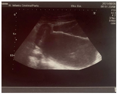
Figure 2 Abdominal ultrasound: regular anteverted uterus with linear endometrium. Anteuterine formation compatible with hematocolpos.
The therapeutic approach consisted of hymenectomy. In the operating room, under general anesthesia a 1 cm longitudinal incision was made at the level of the hymeneal membrane followed by drainage of abundant dark hematic material.
The patient is currently asymptomatic and has regular menstrual cycles, without any adverse event.
Discussion
Imperforate hymen, despite being a rare disease, is the most frequent congenital anomaly of the female internal genitalia, with an incidence of 1 per 1,000 women, according to the American College of Obstetricians and Gynecologists (ACOG)5.
It has been associated with amenorrhea and hematocolpos, being the cause of 90% of hematocolpos, but it is the least frequent among the different causes of amenorrhea. Most occur sporadically, although cases associated with autosomal dominant and recessive inheritance have been described1,2,4.
The absence of hymenal perforation is related to an alteration in the canalization of the vagina and the absence of degeneration of epithelial cells during embryonic development1,2. In addition to imperforation, there are other pathological forms of the hymen such as microperforation, cribriform or septate hymen4,6, being the annular and fimbriated the most common within normal hymens1.
The form of presentation is variable. Most of the symptoms are derived from the accumulation of blood due to the obstruction generated by the imperforate hymen. In the study by Darwish et al3, of the 36 patients evaluated with imperforate hymen, 100% presented hematocolpos and 70% had cyclic pelvic pain.
Although rare, associated anomalies at the level of the kidneys, ureters or bladder should be ruled out, since the development of the genital tract is closely related to the urinary system6.
Lee K. et al7 conducted a study in which 236 women with imperforate hymen were included. 27.7% showed urinary retention, while only 5% had renal involvement. Cases of repeated urinary tract infections and urinary incontinence related to accidental penetration of the urethra have been described3.
In the diagnosis, the importance of a good anamnesis about menarche and coital history is emphasized1. Most are diagnosed between 1218 years of age by examination of the internal genitalia7, visualizing a bulging bluish membrane covering the entire vaginal introitus1. When performing the Credé maneuver, it is possible to observe the increase of the bulge1.
Although they are usually diagnosed at puberty, symptomatic pediatric cases have been detected and even cases diagnosed by prenatal ultrasound after the finding of hydrocolposis1,2. These are generated by accumulation of urogenital secretions under maternal estrogenic influence and most of them end up being reabsorbed6,8.
The importance of not delaying diagnosis lies in the possibility of developing complications such as hydronephrosis, endometriosis and infertility, in addition to perforation of the hematocolpos1. The few published cases of death have been associated with respiratory failure due to abdominal distension1,2,4.
Abdominal ultrasound is useful in the confirmation of the diagnosis, not requiring any additional test9. However, for differential diagnosis, pelvic MRI with anatomical study of the pelvis may be necessary1.
Vaginal agenesis, partial hymenal obstruction and low transverse vaginal septum are included in the differential diagnosis5. The association with McKusick Kaufman syndrome, which also includes polydactyly, congenital heart disease and hydrometrocolpos should be considered1. However, most cases occur in isolation, as demonstrated by Lee K. et al7 who, of the 236 cases studied, 116 had imperforate hymen without other associated pathology.
Partial obstruction of the hymen, which can manifest as septate, cribiform or microperforated hymen, is usually asymptomatic, with consultation due to the impossibility of using a tampon or coital problems. Treatment is surgical, similar to surgery for imperforate hymen, resecting the excess hymenal tissue6.
Vaginal agenesis consists of the absence of vagina with variable development of the uterus. Although less frequent than imperforate hymen, it is the second most frequent cause of primary amenorrhea after gonadal dysgenesis. On physical examination, the external genitalia are normal, presenting a small concavity at the vaginal level and absence of the hymen. Unlike imperforate hymen, the first-choice treatment recommended by ACOG is the use of vaginal dilators, with excellent results and a high degree of patient satisfaction6,8.
Transverse vaginal septum consists of the formation of a septum, in 46% of cases at the level of the upper third of the vagina, partially obstructing the vagina and potentially developing hematocolposis. On examination, the septum prevents visualization of the cervix, requiring the use of ultrasound or MRI to distinguish it from agenesis of the cervix. Treatment is surgical, although with worse results than hymenectomy of imperforate hymen, with a higher rate of recurrences and comorbidities such as dyspareunia and dysmenorrhea6,8.
The treatment of imperforate hymen is surgical. The ACOG recommends cross hymenectomy, which consists of draining the accumulated material through an incision at the level of the hymenal membrane9.
Several studies have compared different types of surgical approaches, evaluating efficacy and risk of recurrence. They include cross hymenectomy, hymenectomy by electric scalpel, use of the Foley catheter and dilatation by Hegar dilators. There were no statistically significant differences in the number of restenosis depending on the type of technique used. However, the use of topical estrogen creams is discouraged due to a slight increase in recurrences after their use1.
Some researchers criticize the Foley catheter technique, since maintaining it for at least two weeks increases the risk of local infection3.
The innovative technique in the study by Darwish et al3 demonstrated a high satisfaction rate for patients with imperforate hymen. The procedure consisted of performing the hymenal incision with the triangular tip of a 10 mm laparoscopic trocar.
One of the main drawbacks of the treatment is related to the patient's religious customs. However, information should be provided on the possible complications of not performing the operation, and there are serious cases in the literature, such as perforation of the hematocolpos with generalized peritonitis1,3.
After surgery, the symptoms reported by the patient disappear, with a very low percentage of recurrences. It is estimated that only 6.6% manifest restenosis or vaginal adhesions after surgery7,8. Even in cases where infection has been reported after the incision, the evolution is satisfactory. Postoperative comorbidity is low and 86% of patients do not present dyspareunia1. It is therefore advisable to carry out a subsequent check-up in order to assess the absence of recurrences and adverse effects.
REFERENCES
1. Abdelrahman HM, Feloney MP. Imperforate Hymen. En: Stat- Pearls [Internet]. Treasure Island (FL): StatPearls Publishing; 2021 [citado 4 de noviembre de 2021]. PMID: 32809411 [ Links ]
2. Bonello K, Tlili Y, Ben Zeid O, Figoni H, Mazeghrane M. Haematocolpos due to imperforate hymen disguised as constipation. J Paediatr Child Health. 2021 May;57(5):715-7. doi: 10.1111/jpc.14931 [ Links ]
3. Darwish AM. A Novel Technique for the Reconstructive Formation of an Annular Hymen in Cases of Postpubertal Imperforate Hymen. Sultan Qaboos Univ Med J. 2021 Feb;21(1):e110-e115. doi: 10.18295/squmj.2021.21.01.015 [ Links ]
4. Jang E, So KA, Kim B, Lee AJ, Kim NR, Yang EJ, Shim SH, Lee SJ, Kim TJ. Delayed diagnosis of imperforate hymen with huge hematocolpometra: A case report. World J Clin Cases. 2021 Oct 16;9(29):8901-5. doi: 10.12998/wjcc.v9.i29.8901 [ Links ]
5. Williams Obstetricia. 26ª edición. Editorial: Mc Graw Hill castellano, 2022, página 38. [ Links ]
6. Marc R Laufer, MD (2023) Congenital anomalies of the hymen and vagina. In: Robert L Barbieri, MDDeborah Levine, Alana Chakrabarti, MD, editors. UpToDate 2023. Disponible en: https://www-uptodate-com.bvcscm.a17.csinet.es/contents/congenital-anomalies-of-the-hymen-and-vagina?search=Treatment%20of%20imperforate%20hymen&source=search_result& selectedTitle=1~18&usage_type=default&display_rank=1 [ Links ]
7. Lee KH, Hong JS, Jung HJ, Jeong HK, Moon SJ, Park WH, Jeong YM, Song SW, Suk Y, Son MJ, Lim JJ, Shin JI. Imperforate Hymen: A Comprehensive Systematic Review. J Clin Med. 2019 Jan 7;8(1):56. doi: 10.3390/jcm8010056 [ Links ]
8. Tanitame K, Tanitame N, Urayama S, Ohtsu K. Congenital anomalies causing hemato/hydrocolpos: imaging findings, treatments, and outcomes. Jpn J Radiol. 2021 Aug;39(8):733-40. doi: 10.1007/s11604-021-01115-7 [ Links ]
9. Diagnosis and Management of Hymenal Variants: ACOG Committee Opinion, Number 780. Obstet Gynecol. 2019 Jun;133(6):e372-e376. doi: 10.1097/AOG.0000000000003283 [ Links ]
Received: February 10, 2023; Accepted: June 16, 2023
 Este es un artículo publicado en acceso abierto bajo una licencia Creative Commons
