


Services on Demand
Journal

Article
 text in
  English (pdf)
 Article in xml format
 Article references
 Send this article by e-mail
Indicators
-
 Cited by SciELO
Related links
-
 Similars in
SciELO
Similars in
SciELO
Share

 Permalink
Revista Peruana de Ginecología y Obstetricia
On-line version ISSN 2304-5132
Rev. peru. ginecol. obstet. vol.69 no.3 Lima July/Sep. 2023 Epub Oct 16, 2023
http://dx.doi.org/10.31403/rpgo.v69i2560
Case reports
Vulvar fibroepithelial polyp: a case series

1. General Physician, School of Medicine. Universidad Peruana Cayetano Heredia.
2. Medical Specialist, Department of Gynecology and Obstetrics. Hospital Edgardo Rebagliati Martins.
3. Medical Specialist, Department of Gynecology and Obstetrics, Hospital San José de Chincha.
Fibroepithelial polyps are common benign skin tumors in the general population. However, genital tract involvement is unusual. Their etiology is unclear, but associations with metabolic disorders and hormonal fluctuations have been described, which explains their higher prevalence in women. Due to the variety of differential diagnoses, histopathological evaluation is necessary. Their management is usually conservative. However, they may require surgical intervention in some cases. We present four cases of vulvar fibroepithelial tumors of different sizes, one of them classified as giant, as well as the management approach. With this presentation, we hope to improve knowledge, diagnostic accuracy and contribute to the effective treatment of patients with this rare vulvar pathology.
Key words: Vulva; Neoplasm; benign; Skin neoplasia
Introduction
The study of vulvar pathology plays a crucial role in the understanding and management of various conditions affecting the female genitalia. The vulva, composed of diverse anatomical structures, can be affected by a wide range of benign and malignant diseases.
Fibroepithelial polyps (FEPs) or acrochordons are benign skin tumors of mesenchymal and ectodermal origin. They occur frequently in the general population, and their frequency increases with age1. These tumors are frequently found in skin folds, such as the neck, axilla, submandibular or inguinal areas1. However, the genital tract can also be affected2.
In this case series, we present three cases of vulvar fibroepithelial tumors of different sizes and their management approach, as well as a review of the literature. With this presentation, we hope to improve knowledge, diagnostic accuracy and contribute to the effective management of patients with this rare vulvar pathology.
Cases report
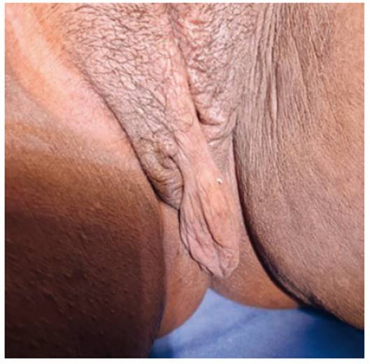
Case 1. A 30-year-old female patient presented for consultation due to a soft pedunculated mass in the right labium majus for the past 12 months (Figure 1). Initially, the mass was approximately 1 × 1 cm in size, which gradually increased, associated with a pulling pain in the perineum. She had no history of pathology or any sexually transmitted infection. Physical examination revealed an irregular mass measuring 20 × 15 × 5 cm arising from the right lip with a 1-cm diameter stalk. It was soft and mobile. The overlying skin was hyperpigmented and with a velvety surface, and there was a pressure ulcer in the lower area. The neck, axilla, groin, and other areas of the body were free of lesions. All physical examination and laboratory findings were normal. Surgical excision of the lesion was performed without incident. Histopathology revealed the diagnosis of benign fibroepithelial polyp. There were no malignant cells in the specimen.

Case 2. A 36-year-old female patient came to the outpatient clinic for a mass on the left vulva, which started as 1x1 in size and increased in size over a ibroepit one year. There was no history of vaginal bleeding, trauma, sexually transmitted diseases, medical or surgical history (Figure 2). Her menstrual history was normal. She used hormonal contraceptives (progesterone injection every three months) for two years. Physical examination was normal. On gynecologic examination, a 7 × 5 × 5 cm, firm, non-painful, pinkish, pedunculated mass protruding from the left labium majus with a pressure ibroepi the anterior ibroep was found. Laboratory results were within normal limits. Surgical excision of the mass was performed. The postoperative course was uneventful. The histopathological ibroepit the biopsy was ibroepitelial polyp of the vulva.

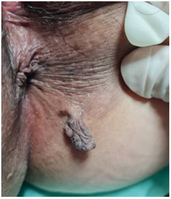
Case 3. A 57-year-old female patient presented with a vulvar mass that increased in size over two years until it reached a size of 5x3x2 (Figure 3). The lesion was asymptomatic except for slight pulling pain on movement. Physical examination revealed a pedunculated mass on the right labium majus covered by normal skin with no signs of ulceration. The rest of the physical examination and laboratory tests were normal. Total surgical resection of the mass was performed, and histopathology of the specimen revealed fibroepithelial polyp.
Case 4. A 45-year-old female patient came to the outpatient clinic for painless mass growth in the perineal area in the last 6 months (Figure 4). She reported no other history. Physical examination revealed a 1 x 1 x 1 pedunculated mass covered by hyperpigmented skin, with no visible ulcers. All physical examination and laboratory findings were normal. Complete excision surgery was performed due to cosmetic issues. The histopathological report showed fibroepithelial polyp.
Discussion
FEP are tumors of frequent occurrence in divers anatomical areas. The first case in the vulva was reported in 1988 by Ostor et al3 and since then few cases have been described in the literature.
These tumors vary in appearance from papules approximately 2 millimeters wide and high also known as acrochordons, filiform lesions approximately 2 millimeters wide and 5 millimeters high, to large pedunculated tumors4. The largest case of vulvar FEP reported in the literature was a lesion 42 centimeters long5. In our cases the variety of sizes of presentation can be evidenced, the largest being 20 centimeters long.
The etiology of FEP has not been completely studied, but an association has been found with the presence of metabolic abnormalities, such as obesity, diabetes mellitus type 2, insulin resistance, dyslipidemias, hypertension, and low high-density lipoproteins (HDLs) levels6. However, none of our patients presented metabolic alterations.
There are different hypotheses about the overgrowth of FEP in the vulvar area. One possible explanation is the sensitivity to hormones and hormonal changes of the epithelium located in the genital tract. Another important feature is that FEPs located in this area are more frequently observed in women of reproductive age, with hormone use and during pregnancy7,8. Understanding the molecular mechanisms of FEP growth could contribute to the development of more specific therapeutic interventions in the future.
The growth rate of fibroepithelial polyps should also be considered. While most patients present slow and steady growth, there are cases in which small lesions exhibit exponential growth in a short period of time9. Some articles even describe de novo onset and rapid growth10, which resembles the first case described.
Due to their variety of clinical presentation, FEP can pose diagnostic challenges because of their overlapping features with other vulvar lesions, such as condylomata or squamous papillomas. In such cases, histopathologic evaluation is necessary.
Histologically, FEP can be of two types according to predominance: epithelial or stromal9. In general, they are composed of a fibrovascular stroma rich in collagen and fibrous tissue, a base with regular boundaries and thin vascular walls that extend parallel to each other. In addition, it is covered by a surface of keratinizing epithelium that can be thick with acanthosis, papillomatosis and hyperkeratosis11.
Although FEP is generally considered a benign and asymptomatic tumor, isolated cases have been reported whose final diagnosis was basal cell carcinoma or squamous cell carcinoma in situ, highlighting the role of histopathology in challenging situations12. In our cases, the definitive diagnosis was confirmed based on histopathologic examination, and none of the specimens evidenced malignant cells.
The management of FEPs is usually conservative; however, they can be treated surgically for esthetic reasons or for chronic irritation. They can be treated with scissors, cautery, or cryotherapy if they are small in size. When they are larger, as in these cases, treatment is performed by total excision under local or regional anesthesia to avoid regrowth. In addition, long-term follow-up is recommended to detect recurrences as early as possible13,14. In the four cases presented, treatment was by total surgical excision using local anesthesia, with adequate healing and complete recovery in all cases.
REFERENCES
1. Marks JG, Miller JJ. Lookingbill and Marks' Principles of Dermatology. Elsevier; 2018. [ Links ]
2. Agrawal A, Garg C, Mukherjee S, Kakkar K. Giant acrochordon of vulva: a rare occurrence. Nepal J Dermatol, Venereol Leprol. 2016;13(1):70-2. doi: 10.3126/njdvl.v13i1.14310 [ Links ]
3. Ostör A, Fortune DW, Riley CB. Fibroepithelial polyps with atypical stromal cells (pseudosarcoma botryoides) of vulva and vagina. A report of 13 cases. Intern J Gynecol Pathol. 1988;7(4):351- 60. doi: 10.1097/00004347-198812000-00006 [ Links ]
4. Elder DE, Elenitsas R, Rubin AI, Ioffreda M, Miller J, Miller OF 3rd, Yun SJ. Atlas of Dermatopathology: Synopsis and Atlas of Lever's Histopathology of the Skin. 4th ed. Lippincott Williams & Wilkins (LWW); 2020. ISBN: 978-1-97-512463-2. [ Links ]
5. Kumar A, Hasin N, Sinha AK, Kumar S, Bhadani P. Giant fibro epithelial polyp in a young girl: A rare case report. Int J Surg Case Rep. 2017;38:83-5. doi: 10.1016/j.ijscr.2017.06.059 [ Links ]
6. Tripathy T, Singh BSTP, Kar BR. Association of Skin Tag with Metabolic Syndrome and its Components: A Case-control Study from Eastern India. Indian Dermatol Online J. 2019 May- Jun;10(3):284-7. doi: 10.4103/idoj.IDOJ_238_18 [ Links ]
7. Yoo J, Je BK, Yeom SK, Park YS, Min KJ, Lee JH. Giant Fibroepithelial Stromal Polyp of the Vulva: Diffusion-Weighted and Conventional Magnetic Resonance Imaging Features and Pathologic Correlation. J Pediatr Adolesc Gynecol. 2019 Feb;32(1):93-7. doi: 10.1016/j.jpag.2018.08.006 [ Links ]
8. Can B, Ozluk A. Y. Giant fibroepithelial polyps: why do they grow excessively? The Med Bull Sisli Etfal Hosp. 2020;54(2):257- 260. doi: 10.14744/SEMB.2018.33603 [ Links ]
9. Dura M, Aktürk H, Sungur G, Alsalamin WOI. A Giant Fibroepithelial Polyp of the Vulva. Cureus. 2023;15(5):e39152. doi:10.7759/cureus.39152 [ Links ]
10. Madueke-Laveaux OS, Gogoi R, Stoner G. Giant fibroepithelial stromal polyp of the vulva: largest case reported. Ann Surg Innov Res. 2013;7:8. https://doi.org/10.1186/1750-1164-7-8 11. Navada MH, Bhat PR, Rao SV, G N. Large fibroepithelial polyp of vulva. Case Rep Dermatol Med. 2011;2011:273181. doi: 10.1155/2011/273181 [ Links ]
11. Navada MH, Bhat PR, Rao SV, G N. Large fibroepithelial polyp of vulva. Case Rep Dermatol Med. 2011;2011:273181. doi:10.1155/2011/273181 [ Links ]
12. Lortscher DN, Sengelmann RD, Allen SB. Acrochordon-like basal cell carcinomas in patients with basal cell nevus syndrome. Dermatol Online J. 2007;13:21 [ Links ]
13. Bernal S, Olivares CV, Ayala MdLM, Cerda FdJ. Fibroepithelial polyp of the vulva (acrochordon): Presentation of a case and review of the literature. Arch Med Actual Tracto Genital Inferior (AMATGI). 2011;III(4):5 [ Links ]
14. Amin A, Amin Z, Al Farsi AR. Septic presentation of a giant fibroepitelial polyp of the vulva. BML Case Reports. 2018;2018, article bcr-2017 doi: 10.1136/bcr-2017-222789 [ Links ]
Received: May 31, 2023; Accepted: August 23, 2023
 Este es un artículo publicado en acceso abierto bajo una licencia Creative Commons
