


Servicios Personalizados
Revista

Articulo
 texto en
  Inglés (pdf)
 Articulo en XML
 Referencias del artículo
 Enviar articulo por email
Indicadores
-
 Citado por SciELO
Links relacionados
-
 Similares en
SciELO
Similares en
SciELO
Compartir

 Permalink
Revista de la Facultad de Medicina Humana
versión impresa ISSN 1814-5469versión On-line ISSN 2308-0531
Rev. Fac. Med. Hum. vol.20 no.2 Lima abr./jun 2020
http://dx.doi.org/10.25176/rfmh.v20i2.2516
Clinical case
Fox - fordyce disease, A case report
1María Auxiliadora General Hospital, Lima-Peru
Fox-Fordyce disease is a non-infectious, infrequent inflammatory dermatosis of apocrine glands, isolated by a pruritic papular rash that usually begins frequently at puberty, and which may involve vulva, armpit and perianal region. It affects women more frequently, approximately in a 9 to 1 ratio, compared to men. In the presentation of the case of a female patient, 53 years old, in consultation of Gynecology-Obstetrics, of the General Maria Auxiliadora Hospital, for presenting for 5 months, papular eruption in vulvar area, associated with intermittent pruritus. A biopsy of the lesion was requested, and it was sent to the Pathological Anatomy Service, receiving a fragment of dark brown tissue, 0.3 x 0.2 x 0.1cm, which was automatically processed, obtaining a histological sheet, in which evidence dilated apocrine glands, which show a thick secretion made up of mucin, in its light. The patient met the histopathological criteria for the diagnosis of Fox - Fordyce disease. Consider the case presentation because of the infrequent nature of this disease.
Key words: Disease; Pruritus Vulvae; Sweat Gland Diseases; Sweat glands (source: MeSH NLM).
INTRODUCTION
Fox-Fordyce disease was first described in 1902 by George Henry Fox and John Addison Fordyce is an uncommon, inflammatory non-infectious dermatoses of apocrine glands1. It is characterized by chronic processes and manifested by very itchy skin-colored papules, in parts of the body where the apocrine glands are concentrated2. Its etiopathogenesis is little known and affects the areas with apocrine glands: the armpits, the groins, the pubic region, the perineum, the labia majora, the areolas and the umbilicus. It occurs mainly in postpubertal women, between 13 and 35 years old, although prepubertal and postmenopause3,4cases have been reported; without racial predilection and with an incidence not yet established2. It mainly affects women in a ratio of 9 to 1; occasionally some cases have been reported in men1Due to emotional or physical stimulation of the apocrine glands, paroxysmal exacerbations are observed. Its treatment is discussed. Different therapeutic regimens have been used with disparate and not always satisfactory results3,4,5.
The cause is an intraepidermal obstruction of the apocrine gland duct. The hypothesis is determined by hormonal and genetic factors, with histopathological criteria that include hyperkeratosis2, obstruction of the follicular infundibulum2, chronic inflammatory changes in the dermis1, and acinar dilation of the apocrine glands1, which show a thick secretion of eosinophilic mucin in lumen1. Treatment is not simple, as some treatment regimens can have annoying side effects and relapses are common2. Other reviews do not specify a definitive treatment1, but describe some treatment options such as combined oral contraceptives to treat and reduce the symptoms of women of childbearing age1. Even Isotretinoin has been used once in the treatment of this condition2, with variable results.
This aim of this study is to present a clinical case of Fordyce-Fox disease. It was decided to present this case because of the rare occurrence of this disease.
CASE PRESENTATION
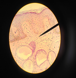
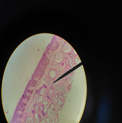
It is presented the case of a postmenopausal 53-year-old female patient, with five years as date of her last period and negative Papanicolaou in 2019, which was evaluated in a gynaecological-obstetric consultation at the General Hospital Maria Auxiliadora, in November 2019, due to a papular eruption in the vulvar region, associated with sporadic itching that she had for 5 months. A biopsy of the lesion was taken and sent to the Pathological Anatomy Service, receiving a dark brown-colored tissue fragment, 0.3 x 0.2 x 0.1 cm, which was properly processed, obtaining a histological sheet, in which dilated apocrine glands were evidenced. Those showed a thick secretion constituted by mucin in lumen; associated with mild inflammatory changes in the dermis (Figure 1y 2). The patient met histopathological criteria for the diagnosis of Fordyce-Fox disease.
It was diagnosed as Fordyce-Fox disease. No medication has proven to be sufficiently effective, so it was decided to keep the patient on a symptomatic treatment.
DISCUSSION
The biopsy of the lesion exposed dilated apocrine glands, which showed a thick secretion of eosinophilic mucin in lumen. Histopathological criteria for the diagnosis of Fordyce-Fox disease include hyperkeratosis2, obstruction of the follicular infundibulum2, chronic inflammatory changes in the dermis1, and acinar dilation of the apocrine glands1, which show a thick eosinophilic secretion of mucin in its lumen1. This case meets two of the four criteria mentioned, which in association with the clinical characteristics of the patient, allowed us to make the final diagnosis of Fordyce-Fox disease, discarding differential diagnoses such as lichen amyloidosis, lichen nitidus, eruptive syringomas, infectious folliculitis, follicular mucinosis, Darier's disease and so on6,7.
Etiology has not yet been clarified. No genetic alterations or associated polymorphisms3have been described; however, Rubio and colleagues involve genetic, endocrine, metabolic or environmental elements with no evidence in favor of any of them3,4,5.
As a first-line therapeutic option, some authors propose the use of calcineurin inhibitors, specifically pimecrolimus. This medication was developed to treat inflammatory processes, such as those characteristic of the disease after the rupture of the glandular duct, and by an unknown mechanism, it inhibits hyperkeratosis and follicular obstruction8. However, other literature does not specify a definitive treatment1; but some treatment options are described, such as combined oral contraceptives to treat and reduce the symptoms of women of childbearing age1, which together with a symptomatic one, is a proposal to start the patient's treatment. The patient received only symptomatic treatment, which was a restriction in the evolution of the clinical condition.
Acknowledgments:
To Dr. Sigrid Abril Santos Laurente, for her teachings and guidance, in this case.
REFERENCES
1. Kurman RJ, Ellenson LH, Ronnet BM. Blaustein´s pathology of the female genital tract. Sixth edition, Springer. [ Links ]
2. Echauri Anahí, Cubilla Elisa, Guzmán Antonio. Enfermedad de Fox-Fordyce: Caso clínico y revisión de la literatura. DermatologíaCMQ2013;11(1):26-28. [ Links ]
3. Rubio C, Mayor M, Marlin MA, González MJ, Contreras F, Gasado M. Enfermedad de Fox Fordyce. Actas Dermosifiliogr. 2004;95:314-6. [ Links ]
4. Consulich Claudia L, Meik S, Vicente AM, Abeldaño A. Pápulas foliculares pruriginosas. Dermatol. 2012;18(5):405-08. [ Links ]
5. García Arpa M, Sánchez Gami Nero P, Vera Iglesias E, Marlin Dávila F. Múltiples pápulas vulvares. Acta Dermosifiliogr. 2007;98(07):499-500. [ Links ]
6. Martínez Luna E, Rebollo Domínguez N, Vega Memije M, Arenas R. Siringomas vulvares: Informe de dos casos. Ginecol Obstet Mex. 2006;74:273-6. [ Links ]
7. Kao PH, Hsu ChK, Lee JY. "Clinicopathological Study of Fox-Fordyce disease". Journal of Dermatology 2009; 36: 485-490.Kao PH, Hsu ChK, Lee JY. "Clinicopathological Study of Fox-Fordyce disease". Journal of Dermatology 2009; 36: 485-490. [ Links ]
8. Chandrakumar A, Francis N, Morar N. "Efficacy of Pimecrolimus in Fox-Fordyce Disease". Open Dermatology Journal 2010; 4: 59-61. [ Links ]
Received: December 08, 2019; Accepted: March 17, 2020
 Este es un artículo publicado en acceso abierto bajo una licencia Creative Commons
