


Servicios Personalizados
Revista

Articulo
 texto en
  Inglés (pdf)
 Articulo en XML
 Referencias del artículo
 Enviar articulo por email
Indicadores
-
 Citado por SciELO
Links relacionados
-
 Similares en
SciELO
Similares en
SciELO
Compartir

 Permalink
Revista de la Facultad de Medicina Humana
versión impresa ISSN 1814-5469versión On-line ISSN 2308-0531
Rev. Fac. Med. Hum. vol.21 no.2 Lima abr-jun 2021
http://dx.doi.org/10.25176/rfmh.v21i2.3621
Clinical case
Patellar ligament rupture as a complication of poor application technique of radial pressure wave therapy. Report of two cases
1Medical Doctor. Orthopedic Surgeon, Centro de Especialidades Ortopédicas, Quito - Ecuador.
2Medical Doctor. Department of Internal Medicine, Centro de Especialidades Ortopédicas, Quito - Ecuador.
3Medical Doctor. Biomedical Sciences Research Institute. Universidad Ricardo Palma, Santiago de Surco - Perú.
4Medical Doctor. School of Medicine, Universidad de El Salvador, San Salvador - El Salvador.
5Medical Doctor. Department of Physical Medicine and Rehabilitation, Hospital General Luis Dávila, Tulcán - Ecuador
6Medical student. School of Medicine, Pontificia Universidad Católica del Ecuador, Quito - Ecuador.
Patellar tendinopathy is characterized by anterior knee pain located at the lower pole of the patella at the junction of the patellar tendon. This is often a disabling condition that limits patients' quality of life, affects their ability to participate in sports, and even hinders their normal daily activities. Extracorporeal shock wave therapy (ESWT) has been recognized as a promising and safe alternative for the treatment of various musculoskeletal disorders - including chronic patellar tendinopathy. However, there is limited evidence regarding its side effects, in particular ESWT-associated tendon injuries. To the authors' knowledge, this is the first report demonstrating clinical and radiological evidence of two patients without known risk factors for partial patellar tendon tears that developed this condition after the application of radial pressure wave therapy - also known as radial shock wave therapy - for patellar tendinopathy. ESWT must be applied by properly trained professionals so that specific requirements needed to guarantee an appropriate application technique, minimize possible adverse effects, and improve patient safety could be met.
Keywords: ligament; Extracorporeal Shockwave Therapy; Tendinopathy; Case reports. SOURCE : Mesh - NLM
INTRODUCTION
Patellar tendinopathy, also called "jumper's knee"1, is characterized by anterior knee pain located at the lower pole of the patella at the junction of the patellar tendon. This condition is common among athletes who perform jumping activities with a prevalence of around 36% in basketball and volleyball players2. More often, patellar tendinopathy is related to injuries produced after repetitive mechanical tension of the patellar tendon producing an inflammatory response with subsequent tendon fiber degeneration. It can limit patients' quality of life, affect their ability to participate in sports, and even hinder their normal daily activities.
On the other hand, patellar ligament rupture is a very rare disorder with an estimated prevalence of 0.6% . Risk factors that may predispose patients to develop this condition include obesity, being high elite or amateur athletes with chronic patellar tendinopathy (generating repetitive microtraumas over the tendon)3, systemic diseases (e.g., rheumatoid arthritis or systemic lupus erythematosus), long-term corticosteroid use, chronic kidney disease, fluoroquinolone use4, elderly, and previous surgical procedures that disturb the midsubstance or insertion sites of the patellar tendon, such as total knee arthroplasty5.
Extracorporeal shock wave therapy (ESWT) has been recognized as a promising and safe alternative for the treatment of various musculoskeletal disorders - including chronic patellar tendinopathy6- due to its practical analgesic effects and ability to promote tissue remodelling and repair6-7.
Currently, there are several shock wave therapy application devices, and each unit varies in terms of the type of wave it provides - radial or focal - thus determining its use for a specific pathology; however, the energy provided by the devices is given primarily on an operator basis, so the risks of applying shock waves do not depend on the device used, but on the protocol established by the operator.
There are currently two wave types, first, a focal shock wave that provides a more significant amount of energy in deep planes generated by piezoelectric, electromagnetic, or electrohydraulic equipment that can activate reparative cellular processes even in bone tissue. Second, a radial pressure wave that is generated through the kinetic transmission of energy on an applicator and has a more superficial effect on the tissues.8
The Latin American Societies and Associations on Shockwave in Medicine (ONLAT)9has made efforts to define the risk factors linked to complications after shock wave therapy application. These include:
Diagnostic errors. Prior to the administration of shock wave therapy, the type of pathology must be correctly identified - e.g. tendinopathy; calcific or fibrous tendinopathy; tendon tear; unspecific pain syndrome.
Technical errors generated by the operator. The operator must be qualified and trained by the corresponding scientific societies. In addition, the anatomy, physiology and pathophysiology of the disease being treated, as well as the indications and contraindications of the therapy, must be completely understood. Lack of knowledge about the type of device and its characteristics is also classified as a technical error. Knowing the correct application technique of shock wave therapy is mandatory, e.g. correct position of the patient during the procedure, localization of the site to be treated -which is generally identified under ultrasonographic guidance-, and establishment of the ideal energy protocol for each pathology.
Adverse effects of the radial wave therapy application such as increased pain, erythema, bruising, among others. Adverse effect management should be addressed by the medical personnel.
There is limited evidence regarding the side effects of the therapy, in particular ESWT-associated tendon injuries. In fact, there is only one case in the literature reporting an Achilles tendon rupture after ESWT10; consequently, to the authors' knowledge, this is the first report demonstrating two cases of partial patellar tendon tears after the application of radial pressure wave therapy also known as radial shock wave therapy.
Case 1:
A 16-year-old previously healthy boy arrived at Centro de Especialidades Ortopédicas in Quito complaining about intense right knee pain. The pain started insidiously three months before when the patient started practicing basketball at his high school. Initially, the patient was evaluated in another medical facility where he was prescribed seven radial pressure wave therapy sessions at a one-week interval with an energy of 5 Bar and a high frequency of 15 to 20 Hz using a BTL-5000 SWT POWER equipment, and no anesthesia was administered. Ultrasonography performed before the therapy demonstrated complete tendon fiber integrity. (Figure 1).
According to the patient, the first radial pressure wave therapy session abruptly increased his knee pain, making him unable to continue walking. As a result, no more ESWT were administered, and one week later he received an intra-articular injection of 80 mg of methylprednisolone without sonographic guidance that failed to improve his pain. He then underwent a complete right knee joint immobilization followed by several physical therapy sessions without improving his symptoms.
Six weeks after receiving the intra-articular corticosteroid injection, the patient was referred to our clinic, where physical examination revealed diffuse swelling on the right anterior knee, significant pain and tenderness over the patellar tendon (9/10 in the Visual Analogue Scale), difficulty on leg extension, and a decreased range of motion. Positive Zohler's and Clarke's tests were present, representing concomitant signs of grade I patellar chondromalacia. The Victorian Institute of Sports Assessment (VISA-P) score for patellar tendinopathy was 38/100, and the patient's Blazina Scale was IIIb.
Blazina Scale11classifies the disease based on five stages which determine the severity of the injury through symptoms that occur at different levels of sport or activity.
Table 1. Blazina Scale
| Classification | Symptoms |
|---|---|
| Stage 0 | No pain |
| Stage I | Pain after intense sports activity |
| Stage II | Pain at the beginning and after sports activity |
| Stage III | Phase III a: Pain during and after activity, but allows regular workouts Phase III b: Pain during and after activity, but unable to perform regular workouts |
| Stage IV | Pain during sporting activity unable to participate in sports to a satisfactory level |
| Stage V | Pain during daily activity. Unable to participate in any sport level |
The VISA-P scale12can be applied to quantify symptoms, function, and the ability to perform sports activities in the context to patellar tendinopathy. In addition, it can be used to monitor the recovery of patellar tendinopathy because it allows an early detection of worsening symptoms. The VISA-P questionnaire consists of 8 items with a score from 0 to 100, and 100 is considered a satisfactory result.
Magnetic resonance imaging of the right knee performed at our clinic revealed a partial patellar tendon tear of 3.8 mm, as well as thickening and edema of its superior portion (Figure 2). In addition, ultrasound elastography demonstrated an intrasubstance patellar tendon tear of 4 mm and calcification of its deep portion with a stiffness value of 5 kPa., whereas the resting normal tendinous fibers demonstrated a stiffness value of 1.2 kPa. (Figure 3).
Conservative management was decided considering the patient's age. First, the administration of two 4ml intratendinous injections of PRO-PRP KIT leukocyte-poor platelet-rich plasma using interventional ultrasound was performed. This was followed by several physical therapy sessions, and administration of two low-energy sessions of focal extracorporeal shock wave therapy (fESWT) at a one-week interval. Weeks after treatment, we documented a complete tendon healing by performing serial ultrasound examinations. The pain almost resolved (1/10 in the Visual Analogue Scale), and his VISA-P score was 88/100, so the patient was able to return to normal activities.
It is important to mention that we decided to apply treatments other than physical therapy, medication, and rest, because this patient had previously been treated with various conservative therapeutic measures that did not show favorable results. In addition, it was also decided to apply these "unconventional" treatments due to the large amount of fibrosis and calcification that the patient presented in the tendon insertion area for which both PRP and the use of focal shock waves have shown benefits. On the other hand, it is necessary to emphasize that this patient’s age absolutely contraindicated the use of high intensity laser due to the ray´s depth of penetration which can affect the patient’s growth plate.
Case 2:
A 25-year-old previously healthy man arrived at Centro de Especialidades Ortopédicas in Quito complaining about intense right knee pain that started insidiously several months ago after starting playing soccer on weekends. Initially, he sought medical attention in another medical facility where twenty radial pressure wave therapy sessions on the right knee were prescribed at a one-day interval with an energy of 4 Bar and a frequency of 10 to 15 Hz using a BTL-6000 SWT EASY equipment, and no anesthesia was administered. Ultrasonography performed before the therapy demonstrated proximal patellar tendon thickness and edema, as well as complete tendon fiber integrity with no signs of peritendinous vascularization. (Figure 4).
After the fifth shockwave therapy session, the patient denoted significant pain increase over the application area, and over the next few days, he was unable to walk or climb stairs, so he decided not to continue receiving the therapy and looked for a second evaluation at our clinic.
The patient's physical examination at our clinic showed a positive Bassett sign and an extremely painful right knee extension (9/10 in the Visual Analogue Scale). Furthermore, edema and ecchymosis were noticed over the patellar region of the right leg. The Victorian Institute of Sports Assessment (VISA-P) score for patellar tendinopathy was 37/100, and the patient's Blazina Scale was IIIb.
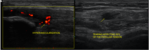
We performed an ultrasound that evidenced a right partial patellar tendon tear compromising approximately 50% of its whole depth surrounded by multiple blood vessels. (Figure 5).
Using interventional ultrasound, we administered 4 intratendinous and 10 peritendinous ml of PRO-PRP KIT leukocyte-poor platelet-rich plasma. Afterward, the patient was advised to use crutches for two weeks followed by administration of ten continuous-mode 5.00-W-power high-intensity laser (HIL) therapy sessions at one-day intervals over the right patellar tendon using a BTL- high intensity laser- 12W equipment.
Finally, twenty physical therapy sessions were prescribed at one-day intervals. The sessions were focused on stretching of the lower extremity musculature, friction massage of the patellar tendon, eccentric quadriceps exercises, and strengthening the hip and knee musculature. The patient achieved a complete tissue and functional recovery, and three months after treatment, his VISA-P score was 91/100.
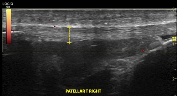
Figure 1. Case 1. Ultrasonography of the right patellar tendon performed before radial pressure wave therapy application demonstrated complete tendon fiber integrity
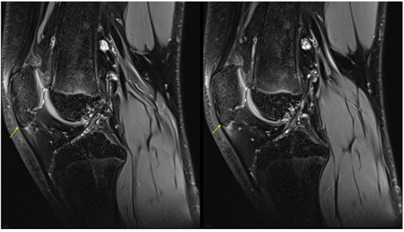
Figure 2. Case 1. Magnetic resonance imaging of the right knee approximately seven weeks after radial pressure wave therapy application revealed a partial patellar tendon tear of 3.8 mm, as well as thickening and edema of its superior portion
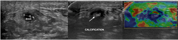
Figure 3. Case 1. Ultrasound elastography performed approximately six weeks after receiving intra-articular knee corticosteroid injection demonstrated an intrasubstance patellar tendon tear of 4 mm and calcification of its deep portion with a stiffness value of 5 kPa
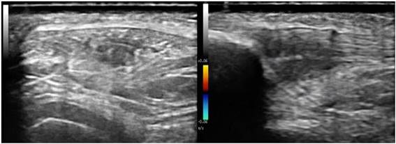
Figure 4 Case 2. Ultrasonography performed before radial pressure wave therapy application demonstrated proximal patellar tendon thickness and edema, as well as complete tendon fiber integrity with no signs of peritendinous vascularization
DISCUSSION
Multiple evidence supports the use of extracorporeal shockwave therapy (ESWT) as effective and safe in treating patellar tendinopathy. Liao et al.,13in a recent meta-analysis of randomized controlled trials evaluating the efficacy of extracorporeal shock wave therapy for knee tendinopathies and other soft tissue disorders, concluded that ESWT exerts a positive effect on the treatment success rate, pain reduction, and range of motion restoration in patients with knee soft tissue disorders. Similarly, Van Leeuwen et al.,7in a review of the literature involving seven studies in which more than two hundred patients with patellar tendinopathy were treated with ESWT, concluded that this treatment seems to be a safe and promising alternative for this tendinopathy with a positive effect on pain and function. Lastly, Wang et al.,14, in a randomized controlled clinical trial evaluating the efficacy of extracorporeal shockwave therapy compared to conservative treatment for chronic patellar tendinopathy, found positive outcomes in 90% of patients in the study group compared to only 50% of patients in the control group. Furthermore, recurrence of symptoms occurred in only 13% of patients in the study group and 50% in the control group.
Concerning the side effects of the therapy, even though Furia et al., have reported some mild side effects as a consequence of ESWT such as ecchymosis, petechiae, slight swelling, and temporary reddening of the skin15, there are very few reports in the literature demonstrating severe complications like the ones seen in our patients.
At date, there is only one previous study reporting a tendon rupture after ESWT10. In this case, a female patient with a history of chronic calcific Achilles tendinopathy experienced Achilles tendon rupture two months after being treated with ESWT. The study concluded that despite ESWT is generally considered safe, physicians should be aware of potentially significant complications such as tendon tears.
It is worth mentioning that none of our patients had any of those risk factors. They also had pre-therapy patellar tendon ultrasonography that demonstrated complete tendon fiber integrity; hence, we can only conclude that their patellar tendon ruptures were produced as iatrogenic consequences of poor application technique of radial pressure wave therapy.
Even though the patients’ histories differ from the ones who develop patellar tendinopathy, it is worth mentioning that the patients’ symptoms started to develop right after a sudden increase in their physical activities - they started to practice a new sport -. This precipitated change in physical activity may have contributed to the development of patellar tendinopathy. In this context, it is well established that progressing physical loading, high intensity training, or repetitive loading too fast may contribute to the development of this condition.16. In addition, it is possible that an interaction between various intrinsic and extrinsic factors with the genetic make up of our patients could have increased their likelihood to develop tendinopathy.17.
Another aspect to consider is that in the first case the patient’s age made the diagnosis of patellar tendinopathy less feasible; however, complementary exams like plain radiographs, MRI and ultrasonography failed to confirm other causes of patellar tendon pain, e.g. Sinding Larsen Johansson Syndrome.
Even though there is enough evidence supporting the use of ESWT for the treatment of patellar tendinopathy, we have identified health providers' lack of expertise as a major risk factor for ESWT side effects18. As mentioned previously, risk factors may play an essential role in developing patellar tendon rupture, but several studies suggest that this condition is often the result of direct trauma over the tendon among healthy individuals such as our patients3. When radial pressure wave therapy is used, tendinopathies are treated with an energy between 4 and 5 Bar, and a low frequency ranging from 6 to 10 Hz.6at a one-week interval. However, in the first case, in addition to inappropriately using a high frequency of 15 to 20 Hz., the patient’s age made the use of radial pressure wave therapy contraindicated. Likewise, in the second case, the therapy was applied erroneously at one-day intervals. In regard to this matter, Leal et al.,6in a review about the use of extracorporeal shock wave therapy for chronic patellar tendinopathy, suggest a maximum of five sessions should be applied for the treatment of this tendinopathy when radial pressure wave therapy is used; thus, in both cases, the protocols were breached.
It is worth mentioning a brief description of the treatments we used in order to help achieve a complete tissue and functional recovery in our patients.
In the first case, we administered two low-energy sessions -0,10mJ/mm2- of focal extracorporeal shock wave therapy (fESWT) at a one-week interval. The use of fESWT is well recognized for providing high-quality energy over tissues leading to activation of reparative cellular processes; thus, promoting tissue repair and neovascularization6-8. In addition, there is far more evidence recommending the use of fESWT to treat patellar tendinopathy compared to radial shock wave therapy (6-8-14). This means that even though an inappropriate shock wave therapy application protocol can lead to severe adverse effects as the ones observed in our patients, the appropriate use of ESWT can definitely lead to positive outcomes.
Regarding the use of poor leukocyte platelet rich plasma, there is a lot of emerging evidence supporting its effectiveness in patellar tendinopathy. Many studies suggest that it promotes tendon healing through the delivery of platelet-derived growth factors and bioactive molecules in hyperphysiologic doses that enhance tissue repair mechanisms.19. In addition, studies which investigated the effects of PRP in vitro and in vivo, demonstrated benefits that include improved cellular remodeling and decreased healing time20.
Finally, in the second case, we administered ten continuous-mode 5.00-W-power high-intensity laser (HIL) therapy sessions at one-day intervals. There is evidence supporting HIL effectiveness in inhibiting pain pathways in the nervous system as well as the production of anti-inflammatory effects by locally stimulating blood and lymph circulation, which leads to reduced edema and increased blood supply. Both effects are produced by the generation of heat in the diseased tissue - an increase of approx. 2°C-3°C- and the mechanical stimulation of nociceptors and other nerve terminals21-22. Consequently, HIL has emerged as a reliable, safe, and effective treatment option in the treatment of various musculoskeletal conditions23.
The main limitation of our report is its retrospective nature. Since the patients received radial shock wave therapy in other medical centers, we were unable to collect valuable information regarding the number of shocks used, or the precise site of radial pressure wave therapy application.
CONCLUSION
This report demonstrates the potentially harmful effects of radial pressure wave therapy for patellar tendinopathy in two otherwise healthy individuals. As stated earlier, none of our patients had risk factors that could have compromised their patellar tendon integrity; therefore, traumatic partial patellar tendon ruptures following a poor application technique of radial shockwave therapy are the most consistent diagnosis.
It is important to mention that we were able to evaluate the patients relatively shortly after they developed their patellar tendon tears, so we could appropriately confirm the diagnosis clinically and radiologically.
As we detailed in a previous report, ESWT must be applied by professionals certified by the International Society for Medical Shockwave Treatment (ISMST) or by the Latin American Societies and Associations on Shockwave in Medicine (ONLAT). Therefore, specific requirements needed to guarantee an appropriate application technique, minimize possible adverse effects, and improve patient safety could be met.
More research is required to clarify the pathophysiological mechanisms involved in tissue injury after ESWT.
More research is required to clarify the pathophysiological mechanisms involved in tissue injury after treatment with radial pressure waves when there is application technique failure.
It is essential to follow the recommendations that the scientific evidence provides in order to establish appropriate treatment protocols for each particular patient. When focal shock wave therapy is used for the treatment of patellar tendinopathy, it is recommended to use an energy between 0.10mJ / mm2 and 0.25mJ / mm2, and and frequency of 4 to 7 Hz with one week intervals - 3 sessions on average-6 13.
REFERENCES
1. Eva LLopis, Mario Padrón. Anterior knee pain. European journal of radiology. 2007; 62: 27-43. [ Links ]
2. Zwerver J, Bredeweg SW, van den Akker-Scheek I. Prevalence of Jumper's knee among nonelite athletes from different sports: a cross-sectional survey. Am J Sports Med. 2011;39(9):1984-1988. doi:10.1177/0363546511413370. [ Links ]
3. Rodrigo Pires e Albuquerque, Juliano Prado, Rafael Hara, Evaldo Ferreira, Leonardo Schiavo, Vincenzo Giordano, Ney Pecegueiro do Amaral, João Mauricio Barretto. Epidemiological study on tendon ruptures of the knee extensor mechanism at a level 1 hospital. Revista brasileira de ortopedia. 2015; 47: 719-723. [ Links ]
4. CPT Daniel J. Stinner , MC USA ; MAJ Justin D. Orr , MC USA † ; MAJ Joseph R. Hsu , MC USA. Fluoroquinolone-Associated Bilateral Patellar Tendon Rupture: A Case Report and Review of the Literature . Military medicine. 2010; 175: 457-459. [ Links ]
5. Yang F, Wang GD, Huang R, Ma H, Zhao XW. Ligament augmentation reconstruction system artificial ligaments in patellar tendon reconstruction - a chronic patellar tendon rupture after multiple operations: A case report. World J Clin Cases. 2020;8(4):831-837. doi:10.12998/wjcc.v8.i4.831 [ Links ]
6. Leal C, Ramon S, Furia J, Fernandez A, Romero L, Hernandez-Sierra L. Current concepts of shockwave therapy in chronic patellar tendinopathy. Int J Surg. 2015;24(Pt B):160-164. doi: 10.1016/j.ijsu.2015.09.066. [ Links ]
7. Leeuwen, M. T., Zwerver, J.,& Akker-Scheek, I.. Extracorporeal Shockwave therapy for patellar tendinopathy: A review of the literature. British journal of sports medicine. September,1, 2008: 163-168. [ Links ]
8. Moya, Daniel MD, Ramón, Silvia MD, PhD; Schaden, Wolfgang MD, Wang, Ching-Jen MD, Guiloff, Leonardo MD, Cheng, Jai-Hong MD. The Role of Extracorporeal Shockwave Treatment in Musculoskeletal Disorders. The Journal of Bont and Join Surgeryt. February, 7, 2018: 251-263. [ Links ]
9. Federación Iberoamericana de Sociedades y Asociacioes de Ondas de choque en Ingenieria Tisular (ONLAT). ONLAT. Ondas de Choque en Medicina La nueva frontera. 2003. Disponible en:https://onlat.net/?page_id=2491. 15 de Febrero del 2021. [ Links ]
10. Lin, Tsung-Ching, Cheng-yuan Lin, Cheng-Liang Chou & Cheng-Ming Chiu. Achilles tendon tear following shock wave therapy for calcific tendinopathy of the Achilles tendon: A case report. Physical therapy in sport: official journal of the Association of Chartered Physiotherapists in Sports Medicine. March,13,2012: 189-92. [ Links ]
11. Vetrano, Mario, et al. Platelet-rich plasma versus focused shock waves in the treatment of jumper’s knee in athletes. The American journal of sports medicine. 2013; 41: 495-803. doi: 10.1177/0363546513475345 [ Links ]
12. Kregel, Jeroen; Van Wilgen, Cornelis Paul; Zwerver, Johannes. Pain assessment in patellar tendinopathy using pain pressure threshold algometry: an observational study. Pain Medicine. 2013; 14: 1769-1775. [ Links ]
13. Liao CD, Xie GM, Tsauo JY, Chen HC, Liou TH. Efficacy of extracorporeal shock wave therapy for knee tendinopathies and other soft tissue disorders: a meta-analysis of randomized controlled trials. BMC Musculoskelet Disord. 2018;19(1):278. Published 2018 Aug 2. doi:10.1186/s12891-018-2204-6. [ Links ]
14. Wang CJ, Ko JY, Chan YS, Weng LH, Hsu SL. Extracorporeal shockwave for chronic patellar tendinopathy. Am J Sports Med . 2007;35(6):972‐978. doi:10.1177/0363546506298109. [ Links ]
15. Furia, John & Rompe, Jan & Cacchio, Angelo & Maffulli, Nicola. Shock wave therapy as a treatment of nonunions, avascular necrosis, and delayed healing of stress fractures. Foot and ankle clinics 2010; 15(4): 651-662. [ Links ]
16. Rutland M, O'Connell D, Brismée JM, Sizer P, Apte G, O'Connell J. Evidence-supported rehabilitation of patellar tendinopathy. N Am J Sports Phys Ther. 2010;5(3):166-178. [ Links ]
17. Merzesh Magra, Nicola Maffulli. Genetic aspects of tendinopathy. ELSIEVER. 2008; 11: 243-247. [ Links ]
18. Paul Terán-Vela, Walter Insuasti-Abarca, Diana Martínez-Asnalema, Tania Platero-Portillo, Sebastián Ramos-Rosas, Sussan Llocclla-Delgado. Ulnar nerve injury after radial extracorporeal shock wave therapy identified with high-resolution ultrasonography: Case Report. Revista de la Facultad de Medicina Humana. 2020; 20: 328-333.DOI 10.25176/RFMH.v20i2.2912 [ Links ]
19. Andriolo L, Altamura SA, Reale D, Candrian C, Zaffagnini S, Filardo G. Nonsurgical Treatments of Patellar Tendinopathy: Multiple Injections of Platelet-Rich Plasma Are a Suitable Option: A Systematic Review and Meta-analysis. Am J Sports Med . 2019;47(4):1001-1018. doi:10.1177/0363546518759674 [ Links ]
20. Vander Doelen T, Jelley W. Non-surgical treatment of patellar tendinopathy: A systematic review of randomized controlled trials. J Sci Med Sport. 2020;23(2):118-124. doi:10.1016/j.jsams.2019.09.008 [ Links ]
21. Sielski Ł, Sutkowy P, Katarzyna PO, et al. The impact of high-intensity laser therapy on oxidative stress, lysosomal enzymes, and protease inhibitor in athletes. Chin J Physiol. 2019;62(6):273-278. doi:10.4103/CJP.CJP_40_19 [ Links ]
22. Elsodany AM, Alayat MSM, Ali MME, Khaprani HM. Long-Term Effect of Pulsed Nd:YAG Laser in the Treatment of Patients with Rotator Cuff Tendinopathy: A Randomized Controlled Trial. Photomed Laser Surg. 2018;36(9):506-513. doi:10.1089/pho.2018.4476 [ Links ]
23. Akkurt E, Kucuksen S, Yılmaz H, Parlak S, Sallı A, Karaca G. Long term effects of high intensity laser therapy in lateral epicondylitis patients. Lasers Med Sci. 2016;31(2):249-253. doi:10.1007/s10103-015-1841-3 [ Links ]
Received: January 02, 2021; Accepted: February 14, 2021
 Este es un artículo publicado en acceso abierto bajo una licencia Creative Commons
