


Serviços Personalizados
Journal

Artigo
 texto em
  Inglês (pdf)
 Artigo em XML
 Referências do artigo
 Enviar este artigo por email
Indicadores
-
 Citado por SciELO
Links relacionados
-
 Similares em
SciELO
Similares em
SciELO
Compartilhar

 Permalink
Revista de la Facultad de Medicina Humana
versão impressa ISSN 1814-5469versão On-line ISSN 2308-0531
Rev. Fac. Med. Hum. vol.23 no.4 Lima out./dez. 2023 Epub 30-Nov-2023
http://dx.doi.org/10.25176/rfmh.v23i4.6110
Clinical case
Sacroiliac Arthromyelitis: Case report
1Hospital Santa Rosa. Lima, Perú
2Instituto de investigaciones en Ciencias Biomédicas de la Universidad Ricardo Palma. Lima, Perú.
3Universidad Federico Villarreal.
Infectious sacroiliitis is a rare condition that is sometimes diagnosed late due to the poor specificity of the symptoms. In addition, imaging techniques such as scintigraphy, tomography, and nuclear magnetic resonance are required to assess the extent and severity of the lesions. If early diagnosis and treatment are made, the clinical evolution is satisfactory and leaves no sequelae.
Clinical case: the case of an 83-year-old female patient is presented, with a history of ischemic cerebrovascular disease, high blood pressure, type 2 diabetes mellitus and urinary tract infection, who presented with moderately intense colic-like abdominal pain, afebrile, and reported that at the time After getting up from his chair he felt weakness in his left side. On admission, hypertensive, febrile with T°: 38°C SatO2: 98% with FiO2: 0.4. On physical examination, there was edema in MMII with pitting (+), distended abdomen, pain in the right flank and left hemiparesis. To the laboratory: hemoglobin 14.8 g/dl, platelets 38,000 μl, leukocytes 18,000 μl.
Keywords: Septic Arthritis; Septic Sacrolileitis (source: MeSH - NLM)
INTRODUCTION
Infectious sacroiliitis (ISI) was first described by Poore in 1878, and since then the literature on this infection has mainly come from case reports and small series of patients.1The incidence of ISI is relatively low, approx. 1%. It represents 2% of all cases.
A case of septic arthritis2,3. This can be due to several different reasons, causes including degenerative diseases, trauma, pregnancy, intravenous drug use, immunosuppressive therapy, hemoglobin disorders, inflammatory diseases and infections such as endocarditis and urinary tract or dermatitis; however, these risk factors may only be present in patients in 55% to 60% of identified cases.(1)Unilateral sacroiliitis should guide the diagnosis of ISI,4although unilateral sacroiliitis is associated with spondyloarthropathies (psoriatic arthritis, early, reactive ankylosing spondylitis), which represents a diagnostic challenge.
Contamination can be the product of bacteremia due to a contiguous infection or direct inoculation, as in the case of joint infiltrations.7
Infections can be caused by pyogenic organisms and tuberculosis.5,8
Nonspecific initial symptoms and variable physical examination findings make diagnosis difficult and are often missed from the outset. The clinical manifestations are different, but the most common pain is in the lower back and buttocks, which worsens when walking. Magnetic resonance imaging (MRI) of the pelvis is the gold standard for diagnosing ISI.6
Long-term antibiotic treatment, lasting more than four weeks, is considered adequate treatment.9
CLINICAL CASE
The case of an 83-year-old female patient with a medical record of cerebrovascular disease, high blood pressure, type 2 diabetes mellitus, and urinary tract infection is presented. The patient suddenly presented, two days before admission, moderately intense colic-like pain in the right flank, weakness of the left side of the body, dysarthria, and intermittent febrile symptoms. During the physical examination on admission, regarding vital signs, it was found: BP: 140/70, T: 38.2°. Regarding the skin and pharynx, edema of the lower limbs and fovea was evident (+). At the respiratory level, the vesicular murmur was decreased in both lung fields. Regarding the CV system, a holosystolic murmur was heard. Furthermore, regarding the abdomen, there was pain on palpation in the hypogastrium and regarding the CNS, there was left hemiparesis.
Regarding the somatic physical examination, a flexion, abduction, and external rotation test (FABERE) was performed, positive, Gaenslen-Mennel test, positive; Log Roll test of the left hip, negative; negative signs of root irritation without palpable inguinal lymphadenopathy.
Laboratory tests revealed: basal glucose 537, urea 170, leukocytosis 18,000, plateletopenia 38,000, fed 3%, segmented 80%, lymphocytes 10%, prothrombin T 17.3. In addition, there was a pathological urine test (proteins 2+, nitrites +, blood +, leukocytes +; regarding urinary sediment: red blood cells at 139.92/uL. and white blood cells at 640.20/uL).
A brain Tomography without contrast was performed, which showed tomographic signs in relation to right fronto-parieto-temporal subacute ischemic stroke, dependent on the territory of the right MCA. Likewise, an EKG was performed with a diagnosis of atrial fibrillation with controlled ventricular response and a complete abdominal ultrasound with a diagnosis of left kidney stones and grade II hydronephrosis.
Due to the persistence of intermittent febrile symptoms with a focus to be determined, serial control laboratory tests were performed after seven days, which revealed basal glucose of 221 mg/dL, urea of 211 mg/dL, CRP of 23.28, and creatinine of 3.30 mg. /dL, leukocytes 13,380 xmm3, platelets 42,000, 4% segmented, 88% segmented and 6% lymphocytes.
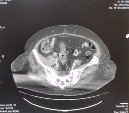
Due to the remarkable symptoms, probable physical examination tests and ultrasound examinations with unilluminating results, it was decided to perform a Tomography of the abdomen with full contrast, with a view to finding sacroiliac disease with retroperitoneal involvement; These images showed at the level of the sacroiliac joint a dense heterogeneous collection with air content associated with cortical erosion that extends anteriorly to the psoas and iliac muscles and posteriorly to the piriformis muscle, as well as tomographic signs in relation to sacroiliac arthromyelitis with areas of abscessed collection in ipsilateral iliac psoas and piriformis muscle (Figure 1)
The diagnosis of retroperitoneal abscess was raised, for which an exploratory laparotomy + abscess drainage + mixed drain was performed. Regarding the operative findings, it is mentioned that upon opening Todd's fascia, bulging of the right psoas was evident due to purulent fluid of approximately 100mL.
DISCUSSION
ISI is a rare disease that presents with non-specific symptoms, often causing a delay in diagnosis. The initial presentation may indicate more common conditions, such as back pain, sciatica, abscesses inside or outside the pelvis, abdominal infection, kidney stones, or pyelonephritis. ISI is usually unilateral. Typical symptoms include low-grade fever, lower back pain (occurring in up to 100% of patients), pain in the waist and back of the thigh, and difficulty walking on the affected side. Surprisingly, more than half of the reported cases were diagnosed 10 days or more after the onset of symptoms2,9.
Early physical examination revealed specific findings, including tenderness in the posterior region of the sacroiliac arthromyelitis and pain caused by compression of the posterior pelvis, but direct palpation of the SIA was difficult, due to its anatomical location, requiring a provocative examination to replicate. pain and symptoms: Gaenslen (forced flexion of the contralateral hip and forced hyperextension of the ipsilateral hip with the patient supine) and a positive FABERE test in up to 91.7% of patients10,11. These tests have been shown to be reliable with sensitivity, specificity, and positive predictive value (60%) in determining the origin of pain, but are not commonly performed in the current clinical setting due to a low index of suspicion.
Berthelot and Laslett12reported that, on clinical examination of SIA, no clinical sign could clearly indicate pain, but that the probability of joint or periarticular pain increases when three of the five most reliable provocation tests are positive:
Iliac wings distraction maneuver
Compression maneuver of the iliac wings
Direct compression on the sacrum
Gaenslen test
Mennell test (Gaenslen test with the patient in the lateral decubitus position)
These tests must be performed on a hard surface, with sufficient duration and force to mobilize the joint and reproduce the pain. The risk of complications, recurrence and sequelae, such as chronic pain, increases with delay in diagnosis.13
Approximately 50% of patients may also have leukocytosis,14but the most reliable laboratory tests are markers of inflammation, including ESR and CRP, and although these markers are sensitive, they are not specific for diagnosis or treatment.3
In the pathophysiology of ISI, infection can be caused by hematogenous dissemination of bacteria from distant sources of ISI; the subchondral circulation on the iliac side of the joint, where the arteries terminate, can serve as an entry point for microorganisms and spread posterior to the joint. Other routes are through adjacent infection, either muscular or intestinal, or by direct inoculation, as in infiltration.7,11
CT can be used for early diagnosis because it shows inflammatory changes and an appearance consistent with ISI. Magnetic resonance imaging is the imaging modality with the highest sensitivity and specificity (95% and 100%, respectively) and is considered the reference test for confirmation of ISI.11MRI combines good visualization of the complex anatomy of Sacroiliac arthromyelitis with the ability to identify varying degrees of inflammation and joint damage, such as the presence of joint and periarticular effusions, bone marrow edema, muscle abscesses, bone erosions and sequestration and Joint capsulitis or widening of the joint space.
Bone marrow edema in sacroiliitis spondyloarthritis occurs mainly in the hip, while in ISI, it occurs mainly in the sacrum or is distributed evenly. 12 On contrast-enhanced MRI, a unilateral increase in uptake is seen three days after symptom onset.6,11,13
The definitive microbiological diagnosis can be based on blood cultures, fluid sampling by percutaneous puncture guided by radiography or CT, or surgical cleansing. Aspiration of the sacroiliac arthromyelitis is technically difficult due to its location.
Under radioscopic guidance, a needle of sufficient caliber is placed to take a tissue sample and aspirate it from the SIJ, to send it for histopathological study, routine cultures, and tuberculosis.14 S. Aureus is the most frequently isolated microorganism, between 45% and 83.3% of cases, according to different authors, followed by coagulase-negative Staphylococcus, group B Streptococcus, Streptococcus pneumoniae, Enterobacteriaceae, such as Escherichia coli, and Salmonella species, mycobacterium catarrhalis, mycobacterium tuberculosis, haemophilus influenzae, brucella species, and pseudomona aeruginosa.2,17,27,28)However, in 27% to 40% of cases, cultures are negative.14
Delay in diagnosis and/or inadequate treatment of IBS can lead to severe consequences, such as bacteremia, septic shock, osteomyelitis or abscess formation.13,14
Currently, there is no consensus on the duration of antibiotic treatment; The usual time is 4 to 6 weeks, although some authors15,16propose 4 to 8 weeks, and others such as Matt et al.17reported that the absence of clinical relapses observed in their group of 18 patients, after a minimum follow-up of 6 months, suggests that 6 to 12 weeks of antibiotic treatment are sufficient to obtain a cure. The choice of antibiotic is based on the culture and the antibiogram.
Surgical intervention is reserved for failure of conservative treatment and the presence of complications such as abscesses and osteomyelitis.18,19
Patient follow-up is prolonged; the absence of symptoms is the first element to consider, in addition to the normalization of inflammatory parameters. Regarding imaging follow-up, it is essential to keep in mind that bone edema persists up to twenty months after completing the treatment.20
CONCLUSION
ISI is a rare disease, but its complications can have serious and functional consequences. A high index of suspicion allows for early diagnosis and rapid initiation of treatment.
Although some diagnoses require the isolation of microorganisms in blood cultures or joint aspirates, the clinical picture of an acute episode, unilateral involvement and intense pain in the buttocks, accompanied by fever, is considered to support the diagnosis of ISI.
Imaging studies, especially MRI, should be performed as soon as possible to aid in early diagnosis. There is no consensus on the duration of antibiotic therapy, but a course of 4 to 6 weeks is most appropriate.
Current information is based on reports and case series, so studies with significant samples are needed to determine the diagnosis, the minimum duration of treatment and the follow-up necessary to control this condition.
REFERENCES
1. Diacinti D, Gioia C, Vullo F, Cannavale G, Catalano C, Valesini G. Magnetic resonance imaging ?ndings of infectious sacroiliitis associated with iliopsoas abscess: a case report in a young male. Reumatismo 2018;70(04):264-267 [ Links ]
2. Kucera T, Brtkova J, Sponer P, et al. Pyogenic sacroiliitis: diagnosis, management and clinical outcome. Skeletal Radiol 2015;44(01): 63-71 [ Links ]
3. Doita M, Yoshiya S, Nabeshima Y, et al. Acute pyogenic sacroiliitis without predisposing conditions. Spine 2003;28(18):E384-E389 [ Links ]
4. Vinceneux P, Rist S, Bosquet A. Arthrites septiques des sacroiliaques et de la symphyse pubienne. Rev Rhum 2006; 73:177-182 [ Links ]
5. Muche B, Bollow M, François RJ, Sieper J, Hamm B, Braun J. Anatomic structures involved in early- and late-stage sacroiliitis in spondylarthritis: a detailed analysis by contrast- enhanced magnetic resonance imaging. Arthritis Rheum 2003;48 (05):1374-1384 [ Links ]
6. Canella C, Schau B, Ribeiro E, Sbaf? B, Marchiori E. MRI in seronegative spondyloarthritis: imaging features and differential diagnosis in the spine and sacroiliac joints. AJR Am J Roentgenol 2013;200(01):149-157 [ Links ]
7. Pertuiset É Les autres causes de sacroiliites que les spondylarthropathies. Rev Rhum 2009;76:761-766 [ Links ]
8. Osman AA, Govender S. Septic sacroiliitis. Clin Orthop Relat Res 1995;(313):214-219 [ Links ]
9. Ghosh S, Narang H, Goel P, Kumar P, Soneja M, Biswas A. Atypical presentation of pyogenic iliopsoas abscess in two cases. Drug Discov Ther 2018;12(01):410 [ Links ]
10. Barnes M, Bush C, Jones J. Pyogenic sacroiliitis: A rare complication of in?ammatory bowel disease. Am J Emerg Med 2019;37(07):1395.e1-1395.e2. Doi: 10.1016/j.ajem.2019.04.017 [ Links ]
11. Wilson JJ, Furukawa M. Evaluation of the patient with hip pain. Am Fam Physician 2014;89(01):27-34 [ Links ]
12. Berthelot J, Laslett M. Par quels signes cliniques s'assurer au mieux qu'une douleur est bien d'origine sacroiliaque. Rev Rhum 2009; 76:741-749 [ Links ]
13. Slobodin G, Rimar D, Boulman N, et al. Acute sacroiliitis. Clin Rheumatol 2016;35(04):851-856 [ Links ]
14. Vyskocil JJ, McIlroy MA, Brennan TA, Wilson FM. Pyogenic infection of the sacroiliac joint. Case reports and review of the literature. Medicine (Baltimore) 1991;70(03):188-197 [ Links ]
15. Hermet M, Minichiello E, Flipo RM, et al. Infectious sacroiliitis: a retrospective, multicentre study of 39 adults. BMC Infect Dis 2012;12:305 [ Links ]
16. Bernard L, Dinh A, Ghout I, et al; Duration of Treatment for Spondylodiscitis (DTS) study group. Antibiotic treatment for 6 weeks versus 12 weeks in patients with pyogenic vertebral osteomyelitis: an open-label, non-inferiority, randomised, controlled trial. Lancet 2015;385(9971):875-882 [ Links ]
17. Scott KR, Rising KL, Conlon LW. Infectious sacroiliitis. J Emerg Med 2014;47(03):e83-e84 [ Links ]
18. Cinar M, Sanal HT, Yilmaz S, et al. Radiological followup of the evolution of in?ammatory process in sacroiliac joint with magnetic resonance imaging: a case with pyogenic sacroiliitis. Case Rep Rheumatol 2012;2012:509136 [ Links ]
19. Sturzenbecher A, Braun J, Paris S, Biedermann T, Hamm B, Bollow M. RM de la artritis séptica. Skeletal Radiol 2000;29:212-215 [ Links ]
20. Shemer A, Eshed I, Levinkopf M. Septic Sacroiliitis: A Diagnostic Challenge for the Clinician. Isr Med Assoc J 2018;20(01):58-59 [ Links ]
Article published by the Journal of the faculty of Human Medicine of the Ricardo Palma University. It is an open access article, distributed under the terms of the Creatvie Commons license: Creative Commons Attribution 4.0 International, CC BY 4.0 (https://creativecommons.org/licenses/by/1.0/), that allows non-commercial use, distribution and reproduction in any medium, provided that the original work is duly cited. For commercial use, please contact revista.medicina@urp.edu.pe.
Received: July 17, 2023; Accepted: October 27, 2023
 Este es un artículo publicado en acceso abierto bajo una licencia Creative Commons
