SciELO - Scientific Electronic Library Online

 
vol.5 número3Aorta shaggy: sustrato ideal para el desastre. Revisión actualizadaTamizaje neonatal de cardiopatías congénitas críticas en el Perú: un llamado de urgencia índice de autoresíndice de materiabúsqueda de artículos
Home Pagelista alfabética de revistas  

Servicios Personalizados

Revista

Articulo

Indicadores

  • No hay articulos citadosCitado por SciELO

Links relacionados

  • No hay articulos similaresSimilares en SciELO

Compartir


Archivos peruanos de cardiología y cirugía cardiovascular

versión On-line ISSN 2708-7212

Arch Peru Cardiol Cir Cardiovasc vol.5 no.3 Lima jul./set. 2024  Epub 29-Sep-2024

http://dx.doi.org/10.47487/apcyccv.v5i3.413 

Special items

Killip and Kimball classification in the Ultrasound era: Is it time to redefine?

La clasificación de Killip y Kimball en la era del ultrasonido: ¿es hora de redefinir?

Marco Antonio Ponce-Gallegos1  *  , Medical doctor
http://orcid.org/0000-0003-4992-9913

Miguel Mendoza-Mujica2  , Medical doctor

Jaime Ponce-Gallegos3  , Medical doctor
http://orcid.org/0000-0002-5389-2028

Jesús Alberto García-Diaz4  , Medical doctor
http://orcid.org/0000-0002-3394-2286

Jorge Armando Zelada-Pineda1 

Diego Araiza-Garaygordobil5  , Medical doctor
http://orcid.org/0000-0003-3185-0051

1Clinical Cardiology Department. Instituto Nacional de Cardiología Ignacio Chávez. Tlalpan, Ciudad de México, México.

2Internal Medicine Department. Nuevo Hospital Civil de Guadalajara “Dr. Juan I. Menchaca”. Guadalajara, Jalisco, México.

3Unidad Cardiológica de Alta Especialidad «Korazón». Tepic, Nayarit, México.

4Clinical Cardiology Department. Hospital Regional “Dr. Valentín Gómez Farías”. Guadalajara, Jalisco, México.

5Coronary Care Unit. Instituto Nacional de Cardiología Ignacio Chávez. Tlalpan, Ciudad de México, México.

ABSTRACT

Lung ultrasound is a tool that is increasingly gaining strength in the initial evaluation of the patient in the emergency department and in critical care areas, making it particularly useful for cardiologists. In patients with ST elevation and acute myocardial infarction it has been observed that 25-45% of patients are wrongly classified as Class I in the Killip and Kimball classification after lung ultrasound (subclinical congestion). The clinical relevance of this finding lies in the fact that the greater the number of B lines, the greater short- and long-term the mortality is. An important advantage is that no prolonged time for learning the technique is required. More studies are needed to evaluate the role and importance of subclinical congestion in patients with acute myocardial infarction. Unfortunately, ultrasound is not widely available in developing countries, so the physical examination will continue to play an important role in the initial evaluation of patients with acute myocardial infraction.

Keywords: Killip and Kimball; Ultrasonics; Myocardial Infarction

RESUMEN

El ultrasonido pulmonar es una herramienta que va cobrando más fuerza en la evaluación inicial del paciente en urgencias y en áreas de cuidados críticos, siendo de especial utilidad para el cardiólogo. Se ha observado que entre el 25 y el 45% de los pacientes con infarto agudo de miocardio con elevación del segmento ST se clasifican erróneamente como Clase 1 en la escala de Killip y Kimball después del ultrasonido pulmonar (congestión subclínica). La relevancia clínica de este hallazgo radica en que cuanto mayor es el número de líneas B, mayor es la mortalidad a corto y largo plazo. Una ventaja importante de esta herramienta es que no se requiere un tiempo prolongado para aprender la técnica. Se necesitan más estudios para evaluar el papel y la importancia de la congestión subclínica en pacientes con infarto agudo de miocardio. Desafortunadamente, el ultrasonido no está ampliamente disponible en los países en desarrollo, por lo que el examen físico seguirá desempeñando un papel importante para la evaluación inicial en pacientes con infarto agudo de miocardio.

Palabras clave: Killip y Kimball; Ultrasonido; Infarto del Miocardio

Since the beginning of the last century, the stethoscope has been one of the most important diagnostic tools for cardiovascular evaluation and diagnosis 1. It is well known that just 50 years ago, the management of practically any condition was based on the data obtained after a complete medical history and physical examination. However, despite the physical examination continues to be a useful resource, available at the patient’s bedside and that can provide a large amount of meaningful data, in numerous settings the advent of new diagnostic modalities, especially ultrasound imaging, have demonstrated a better performance, particularly in cardiovascular physical examination 2,3.

Auscultation plays a key role in the daily work of Cardiologist for detecting evident heart abnormalities, and this pillar of the physical examination has been used as part of some of the most important classifications and scales in Cardiology, such as Killip and Kimball (KK) in the context of acute myocardial infarction (AMI), which was described for the first time in 1967, and has been validated in multiple studies and used widely so far as a prognostic tool. Stages KK I (no signs of congestion) and KK II (heart failure signs [HF], S3 sound and basal rales) have the best prognosis. On the other hand, stages KK III (acute pulmonary edema) and KK IV (cardiogenic shock) confer the worst prognosis at 30 days post-acute myocardial infarction 4,5. Stages III and IV are the most affordable to identify with auscultation. However, mainly stage II can go unnoticed by inexperienced ears.

Given this situation, lung ultrasound (LUS) has come to solve this problem by being able to evaluate pulmonary congestion more objectively. LUS is a focused technique that is fast, and harmless for the patient, and easy to reproduce for the operator and has begun to form part of the cardiologist’s tools, particularly in the emergency department 6. LUS has been validated mainly in patients with acute decompensated heart failure (ADHF). In the LUS-HF trial, it was shown that a LUS-guided diuretic treatment strategy was associated with reduced decompensations and improved walking capacity at six months of follow-up 7. In addition, in a post-hoc analysis of this study, it was demonstrated that subclinical congestion at discharge (defined as the presence of ≥5 B-lines in the LUS test in the absence of rales in the auscultation) was associated with a worse prognosis at 6-month follow-up (mainly by a higher HF admission rate in the group of patients with subclinical pulmonary congestion) with a tendency for higher rates of mortality. Interestingly, outcome rates between patients with subclinical pulmonary congestion and patients with clinical congestion (rales) at discharge were similar 8, which tells us about the great impact that subclinical congestion has on the prognosis of these patients.

In the setting of AMI, there are recent studies that have evaluated pulmonary congestion by LUS in this group of patients. For example, Araujo and co-workers 9 described that in their cohort of patients with ST elevation myocardial infarction (STEMI), 45 patients (41%) who were classified as KK I when performing LUS were classified as “wet lungs” (more than 3-b lines at least in one zone). In the same line, He et al.10, Araiza-Garaygordobil and co-workers 11, and a sub-analysis of the PARADISE-MI trial 12) described a non-negligible number of patients with STEMI that, despite having been classified as KK I had an important number of B-lines in LUS (42%, 42.8%, and 25%, respectively). However, what is the clinical importance of this discordance? Is it clinically relevant? Does it have a real impact on the prognosis of patients? The answer is yes. In all these studies, the presence of B-lines in STEMI patients increases mortality exponentially (the greater the number of B lines, the greater the mortality is) in the short (30 days) and long term (8 months), independently of the KK grade. (Figure 1).

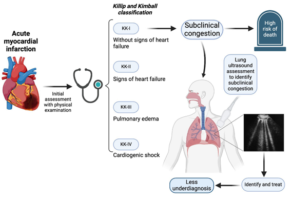
Figure 1 Utility of lung ultrasound in the context of STEMI, mainly in the identification of subclinical congestion in those patients classified as Killip and Kimball Class I. 

Despite growing evidence of the great usefulness of ultrasound as an initial evaluation of STEMI patients, little is known about the clinical impact of the presence of pulmonary congestion in patients classified as KK-I at admission (theoretically there are no signs or symptoms of congestion). A Spanish group directed by Carreras-Mora 13 evaluated 312 patients with STEMI which were classified as KK-I, later dividing them into two groups, wet and dry lung, according to the number of B-lines (3 or more in at least one zone). The presence of early subclinical pulmonary congestion identified by LUS in patients with KK-I STEMI at hospital admission was associated with adverse outcomes during hospitalization and 30-day follow-up. However, it is important to note that the results were bordering.

Araujo et al.9 were the first group that developed a classification that combine clinical and LUS findings named lung ultrasound combined with KK (LUCK I: zero positive zones; LUCK II: either 1) mild B profile or 2) patients with Killip I and severe B profile; LUCK III: either 1) Killip II classification with severe B profile or 2) Killip III patients; and LUCK IV: those with clinical evidence of cardiogenic shock regardless of LUS findings; they demonstrated that LUS added to KK classification was more sensitive than physical examination only to identify patients at risk for in-hospital mortality, therefore, it is necessary to validate this classification with larger population samples, since it could be applicable in any center that has ultrasound in the Emergency Department. In agreement with this study, Dávila-Román and Checkley commented that the addition of LUS to the clinical assessment of critically ill patients provides invaluable prognostic information, coupled with the speed of obtaining it 14. Also, He et al.10) demonstrated that the addition of LUS to the KK classification provides significant incremental prognostic value, which can be an independent predictor of worsening heart failure in AMI during hospitalization and short-term follow-up.

More recently, Carreras-Mora et al.15 created a new simplified classification called “Killip pLUS”, making a comparison between their new classification and the KK and LUCK classifications. This multi-center cohort included 373 patients with the diagnosis of STEMI, and lung ultrasound was performed within the first 24 hours of admission. The authors demonstrated a better performance to predict in-hospital mortality with Killip pLUS (AUC 0.90, 95% CI 0.85-0.95) compared with KK (AUC 0.85, 95% CI 0.73-0.96) and LUCK (AUC 0.83, 95% CI 0.70-0.95). This study demonstrates that the addition of lung ultrasound provides substantial benefits when it comes to predicting worse outcomes in comparison to the exclusive use of the stethoscope.

One of the most important advantages of LUS is that, throughout multiple studies, it has been demonstrated that many hours of training are not necessary to learn how to do it properly (2 hours of reading and 10 ultrasound scans), if there is an expert to supervise, direct, and teach initially 16,17. Iminashi et al.18) addressed this aspect in patients with residual congestion due to heart failure, and they found a substantial agreement inter-operator between residents and the expert (κ = 0.86), as well as a very high (0.91) Spearman rank correlation coefficient for the B-lines between the expert and residents.

Unfortunately, ultrasound devices are not widely available, particularly in developing countries, hence physical examination continues to be of great importance. The contemporary clinic needs to be founded on the experience collected through the centuries and combine it with the technological advances. Despite this, it should not be assumed that new technologies will completely replace the usual methods, since the physical examination will always have an essential role in the decision-making 6. Quoting Dr. Ignacio Chávez: “Medicine was clinical at birth, and it will always be clinical, no matter how profound its scientific transformation. Otherwise, it wouldn’t be medicine”. However, the limitations of the clinic must be considered and know how to use the tools that are available. In this sense, particularly in STEMI, LUS should (or must) be a key tool for the cardiologist and physicians in the ED at the primary evaluation. However, more studies that evaluate subclinical congestion are required to completely establish the role of LUS in STEMI initial evaluation.

Acknowledgments

The authors thank the various participating institutions for the facilities to carry out this manuscript

References

1. Tavel ME. Cardiac auscultation: A glorious past - But does it have a future? Circulation. 1996;93(6):1250-3. doi: 10.1161/01.cir.93.6.1250. [ Links ]

2. Jaffe WM, Roche AHG, Coverdale HA, Mcalister HF, Ormiston JA, Greene ER. Clinical Evaluation Versus Doppler Echocardiography in the Quantitative Assessment of Valvular Heart Disease. Circulation. 1988;78(2):267-75. doi: 10.1161/01.cir.78.2.267. [ Links ]

3. Araiza-Garaygordobil D, Illescas-González E, Cossío-Aranda JE, Kuri-Alfaro J, Guadalajara-Boo JF. Clinical value of cardiovascular physical examination: A review of evidence. Arch Cardiol Mex. 2017;87(4):265-9. [ Links ]

4. Killip 3rd T, Kimball JT. Treatment of myocardial infarction in a coronary care unit. Am J Cardiol. 1967;20(4):457-64. [ Links ]

5. Khot UN, Jia G, Moliterno DJ, Lincoff AM, Khot MB, Harrington RA, et al. Prognostic Importance of Physical Examination for Heart Failure in Non-ST-Elevation Acute Coronary Syndromes The Enduring Value of Killip Classification. JAMA. 2003;290(16):2174-81. doi: 10.1001/jama.290.16.2174. [ Links ]

6. Gopar-Nieto R, Alanís-Estrada GP, Ronquillo-Ramírez DE, Vargas-Estrada JL, Arias-Mendoza A, Rojas-Velasco G, et al. Lung ultrasound in cardiology: Realities and promises. Arch Cardiol Mex. 2019;89(4):369-75. doi: 10.24875/ACM.19000178. [ Links ]

7. Rivas-Lasarte M, Álvarez-García J, Fernández-Martínez J, Maestro A, López-López L, Solé-González E, et al. Lung ultrasound-guided treatment in ambulatory patients with heart failure: a randomized controlled clinical trial (LUS-HF study). Eur J Heart Fail. 2019;21(12):1605-13. doi: 10.1002/ejhf.1604. [ Links ]

8. Rivas-Lasarte M, Maestro A, Fernández-Martínez J, López-López L, Solé-González E, Vives-Borrás M, et al. Prevalence and prognostic impact of subclinical pulmonary congestion at discharge in patients with acute heart failure. ESC Heart Fail. 2020;7(5):2621-2628. doi: 10.1002/ehf2.12842 [ Links ]

9. Araujo GN, Silveira AD, Scolari FL, Custodio JL, Marques FP, Beltrame R, et al. Admission Bedside Lung Ultrasound Reclassifies Mortality Prediction in Patients With ST-Segment-Elevation Myocardial Infarction. Circ Cardiovasc Imaging. 2020;13(6):E010269. doi: 10.1161/CIRCIMAGING.119.010269. [ Links ]

10. He J, Yi S, Zhou Y, Hu X, Lun Z, Dong H, et al. B-Lines by Lung Ultrasound Can Predict Worsening Heart Failure in Acute Myocardial Infarction During Hospitalization and Short-Term Follow-Up. Front Cardiovasc Med. 2022;9:895133. doi: 10.3389/fcvm.2022.895133. [ Links ]

11. Araiza-Garaygordobil D, Baeza-Herrera LA, Gopar-Nieto R, Solis-Jimenez F, Cabello-López A, Martinez-Amezcua P, et al. Pulmonary Congestion Assessed by Lung Ultrasound and Cardiovascular Outcomes in Patients With ST-Elevation Myocardial Infarction. Front Physiol. 2022;13:881626. doi: 10.3389/fphys.2022.881626. [ Links ]

12. Platz E, Claggett B, Jering KS, Kovacs A, Cikes M, Winzer EB, et al. Trajectory and correlates of pulmonary congestion by lung ultrasound in patients with acute myocardial infarction: insights from PARADISE-MI. Eur Heart J Acute Cardiovasc Care. 2023;12(3):155-64. doi: 10.1093/ehjacc/zuad001. [ Links ]

13. Carreras-Mora J, Simón-Ramón C, Vidal-Burdeus M, Rodríguez-Sotelo L, Sionis A, Giralt-Borrell T, et al. Subclinical congestion assessed by lung ultrasound in ST segment elevation myocardial infarction. Heart. 2023;109(21):1602-7. doi: 10.1093/ehjacc/zuad001. [ Links ]

14. Dávila-Román VG, Checkley W. Lung Ultrasound in Cardiac Intensive Care: More Than Just LUCK? Circ Cardiovasc Imaging. 2020;13(6):e010909. doi: 10.1161/CIRCIMAGING.120.010909. [ Links ]

15. Carreras-Mora J, Vidal-Burdeus M, Rodríguez-González C, Simón-Ramón C, Rodríguez-Sotelo L, Sionis A, et al. Killip scale reclassification according to lung ultrasound: Killip pLUS. Eur Heart J Acute Cardiovasc Care. 2024;13(7):566-569. doi: 10.1093/ehjacc/zuae073. [ Links ]

16. Mozzini C, Fratta Pasini AM, Garbin U, Cominacini L. Lung ultrasound in internal medicine: training and clinical practice. Crit Ultrasound J. 2016;8(1):10. doi: 10.1186/s13089-016-0048-6. [ Links ]

17. Parras JI, Onocko M, Traviesa LM, Fernández EG, Morel PM, Cristaldo NG, et al. Lung ultrasound in acute myocardial infarction. Updating Killip & Kimball. Indian Heart J. 2021;73(1):104-108. doi: 10.1016/j.ihj.2020.11.148. [ Links ]

18. Imanishi J, Iwasaki M, Ujiro S, Nakano T, Yamashita T, Eto H, et al. Accuracy of lung ultrasound examinations of residual congestion performed by novice residents in patients with acute heart failure. Int J Cardiol. 2024;395:131446. doi: 10.1016/j.ijcard.2023.131446 [ Links ]

Funding: None declared

Cite as: Ponce-Gallegos MA, Mendoza-Mujica M, Ponce-Gallegos J, García-Diaz JA, Zelada-Pineda JA, Araiza-Garaygordobil D. Killip and Kimball classification in the Ultrasound era: Is it time to redefine?. Arch Peru Cardiol Cir Cardiovasc. 2024;5(3):153-156. doi: 10.47487/apcyccv.v5i3.413

Received: July 07, 2024; Accepted: September 02, 2024

*Correspondence: Marco Antonio Ponce-Gallegos. Juan Badiano 1, Belisario Domínguez Sección 16, Tlalpan, 14080 Ciudad de México, México.+52 3111419479 Email m.antonio.p.g@hotmail.com

Author Contributions: MAPG: Conceptualization and writing original draft; MMM: Writing, review and editing; JPG: Data curation and conceptualization; JAGD: Writing original draft; JAZP: Investigation; DAG: Supervision and project administration

Conflicts of interest: The authors declare that they have no conflict of interest

Creative Commons License This is an open-access article distributed under the terms of the Creative Commons Attribution License